Just because you can …

With a couple of new papers landing that touch on the issue of how you provide and measure quality care around airway management, Dr Alan Garner returns to point at big animals that are bad at hiding.

Two new airway papers have come across my desk in the last couple of weeks and I now wish I had waited a bit longer before putting up the last post on first look intubation as a quality measure.

So where to start? Well how about a place where everything is apparently big? Yes, there’s a bit of work just out of Texas which sheds further light on that first look intubation story so that’s where we’ll land.

Chasing Quality

It sounds like they have used RSI for a while but undertook a quality improvement project to try and reduce their peri-intubation hypoxia rate.  The project involved introducing a bundle of interventions described in the paper as “patient positioning, apn[o]eic oxygenation, delayed sequence intubation, and goal-directed preoxygenation”.

The paper provides copies of the protocol for intubation pre- and post-bundle intervention in the on-line appendices so I might just go through them here to see what they did differently.

The first thing is there was an emphasis on positioning in the bundle, specifically head up a bit and ear-sternum positioning.  Lots of goodness here that I strongly support.

The second measure they mention was apnoeic oxygenation.  However looking at the pre- and post-bundle policies it is evident that they used it in both time periods.  In the before period it ran at 6L/min till the sedation was given then it was turned up to 15L/min.  In the post period however it was run at “MAX regulator flow” after the ketamine was administered.  I don’t know about the O2 regulators in Texas but to me this does not sound like they changed anything significant.  I will come back to apnoeic oxygenation later.

For pre-oxygenation in the pre- bundle period they used a NRB mask (with nasal prong O2 as above) in spontaneously ventilating patients (and arrested patients were excluded) but in the post- period the pre-oxygenation had to be by BVM with two handed technique to ensure a tight seal plus PEEP.  More goodness here that warms my heart.

Delayed sequence intubation in this study refers to administering 2mg/kg of ketamine then maximising preoxygenation for at least 3mins prior to administration of the muscle relaxant.  I don’t think this is necessary in all patients but this was the policy in the bundle.

The last thing they did was “goal-directed preoxygenation”.  This refers to having a SpO2 target >93% for at least 3 minutes during the pre-oxygenation phase after the ketamine had been administered.  If they could not achieve >93% the patient was managed with an LMA or BVM and transported.  I think this represents sensible patient selection in that it removes the high risk of desaturation patients from the process.  When you look at the results you need to keep this patient selection in mind. However I agree that in their system this is a reasonable approach to ensure patient safety for which the managers should be applauded.

Show Me The Money

Yes let’s get to that money shot:

Table

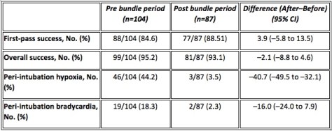
I have been banging on about peri-intubation hypoxia being far more important than first look intubation rate for a while now and this data shows really clearly why.

There is no significant difference in this study in either first look or overall success rates pre and post the bundle but the hypoxia rate fell by a massive absolute 41%!  The 16% decrease in bradycardia emphasises just how much difference they made.  The managers of this system and their staff alike both need to be congratulated for this achievement as this is something that really matters.  And the first pass and overall success rates give no clue!

It really is time to drop first look as a quality measure and move on.  You could look at this paper and start wondering if it might even be worth dropping overall success rate too, which is an interesting thought.  Their policy favoured patient safety over procedural success rates by abandoning the attempt if the pre-oxygenation saturations could not be raised above 93%. It looks like it is working out well for the patients.

Oh, Back to Oxygenation

I promised I would come back to the apnoeic oxygenation issue.  I know the authors state that it was part of their bundle, but it was used in the pre- bundle period as well.  Hence there is no data here to support it’s use.

All three randomised controlled trials of apoeic oxygenation in the ED and ICU contexts (see the notes at the end) have now failed to find even a suggestion that it helps (check those notes at the end for links) and there are no prehospital RCTs.  My take is that it is time to move on from this one too and simply emphasise good pre-oxygenation and good process when the sats start to fall – or never rise in the first place like this group did so well.

Overall a big well done to the Williamson County EMS folks and thanks for sharing your journey with us.

Moving Right Along

The other paper comes out of London, where the ever-industrious HEMS group have published a retrospective review of their database over a 5 year period (from 2009-2014). They were looking for adult trauma patients they reached with an initial noninvasive systolic blood pressure of 90 mmHg or less (or where a definite reading wasn’t there, those with a central pulse only) and with a GCS of 13-15.

This gave them a total of 265 patients (out of a potential 9480 they attended). 118 of those underwent induction of anaesthesia out there beyond the hospital doors (though with exclusions in analysis they end up with 101 to look at) and the other 147 (that number dropped to 135 on the analysis) got to hospital without that happening.

Now the stated indications for anaesthesia listed are actual or impending airway compromise, ventilatory failure, unconsciousness, humanitarian need, patients unmanageable or severely agitated after head injury, and anticipated clinical course.

Now given that the inclusion criteria includes patients having a GCS of 13-15, it seems like both unconsciousness and those really impossible to handle after head injury are likely to be pretty small numbers in that 101. Even airway compromise, ventilatory failure and humanitarian need seem like they’d be not the commonest indications in that list that would apply to this patient group, though they’d account for some.

I guess it’s possible the patients were all initially GCS 13-15 on the team’s arrival but deteriorated en route, though I just can’t sift that out from the paper. Plus if that was the case it seems like you’d say that.

The Outcomes

In their 236 study patients, 21 died and 15 of those were in the ‘received an anaesthetic’ group. The unadjusted odds ratio for death was 3.73 (1.3-12.21; P = 0.01). When adjusted for age, injury mechanics, heart rate and hypovolaemia the odds ratio remained at 3.07 (1.03-9.14; p = 0.04).

Yikes, sort of.

What To Make of That? 

I guess we should make of it that … things you’d expect to happen, happen? Intubating hypotensive patients and then adding positive pressure ventilation in the prehospital setting is potentially risky for patients for a variety of known pharmacological and physiological reasons that the authors actually go into.

So the question is why embark on such a procedure where you know the dangers in detail? You’ve have to really believe in it to end up wiht 101 cases to follow up.

It feels like there’s an elephant in the room to try and address by name. I wonder if it has something to do with a practice I observed while working in the south-east of England 8 years ago. It relates to that last category “anticipated clinical course”.

james-hammond-347179-unsplash
Hovering elephant heads. They’re real.

The concept here is that if you figure the patient is going to be intubated later on in the hospital, you might as well get on and do it. Except the data here suggests that, much like you’d expect, you probably shouldn’t get on and channel your inner Nike marketing script.

Just because you can does not mean you should.  This paper really drives this home though it doesn’t really seem to come straight out and say it. It does pass the comment that “Emergency anaesthesia performed in-hospital for patients with cardiovascular compromise is often delayed until the patient is in theatre and the surgeon is ready to proceed.” Perhaps the problem isn’t using the phrase “anticipated clinical course”. It might be that you just have to remember that the anticipated course might best contain ‘risky things should probably happen in the safest spot’ in the script.

Compare and Contrast

The process of undertaking emergency anaesthesia because later the patient might require emergency anaesthesia is pretty much the complete opposite of the approach from the Williamson County EMS folks. They erred on the side of patient safety and withheld intubation if it was associated with unacceptable risk.

This paper demonstrates that emergency anaesthesia in patients with a high GCS but haemodynamic instability is associated with higher mortality.  We should probably be glad the authors have made this so apparent, because this is probably as good as we’re going to get. We’re not going to get a randomised controlled trial to compare groups. No one is allowing that randomisation any time soon making this another example of needing to accept non-RCT research as the best we’ll get to inform our thinking.

Patients with hypovolaemia due to bleeding need haemorrhage control. The highest priority in patients with that sort of hypovolaemia would seem to be getting them to the point of haemorrhage control quicker. And delaying access to haemorrhage control (because the prehospital anaesthesia bit does add time in the prehospital setting) when the patient has a GCS of 13-15 doesn’t seem to prioritise patient safety enough. Patients probably need us to adjust our thinking on this one.

That seems like common sense. The retrospective look back tells us pretty conclusively it’s a worse option for patients. And now it’s up to us to look forwards to how we’ll view those indications for our next patients. And “anticipated clinical course” probably just doesn’t cut it.


Notes:

That hovering elephant head was posted by James Hammond in a Creative Commons-like fashion on unsplash.com and is unchanged here.

How about all those things that got a mention above that you should really go and read for yourself?

Here’s that whole bundle of care paper out of Texas:

Jarvis JL, Gonzales J, Johns D, Sager L. Implementation of a Clinical Bundle to Reduce Out-of-Hospital Peri-intubation Hypoxia. Ann Emerg Med. 2018;doi:10.116/j.annemergmed.2018.01.044 [Epub ahead of print]

Those RCTs of apnoeic oxygenation in critical care environments mentioned are these ones:

Caputo N, Azan B, Domingues R, et al. Emergency Department use of Apnoeic Oxygenation Versus Usual Care During Rapid Sequence Intubation: A Randomized Controlled Trial (The ENDAO Trial). Acad Emerg Med. 2017;24:1387-1394.

Semler MW, Janz DR, Lentz RJ, et al. Randomized Trial of Apnoeic Oxygenation during Endotracheal Intubation of the Critically Ill. Am J Respir Crit Care Care Med. 2016;193:273-80.  

Vourc’h M, Asfar P, Volteau C, et al. High-flow nasal cannula oxygen during endotracheal intubation in hypoxemic patients: a randomised clinical trial. Intensive Care Med. 2015;41:1538-48.

And that paper on the hypotensive, awake prehospital patients scoring an anaesthetic is this one:

Crewdson K, Rehn M, Brohi K, Lockey DJ. Pre-hospital emergency anaesthesia in awake hypotensive trauma patients: beneficial or detrimental? Acta Anaesthesiol. Scand. 2018;62:504-14.


The Deal with Seals

Greg Brown returns to look at an important thing relevant to first responders (and lots of other people really) – the sucking chest wound. 

We’ve all been there – sitting through some kind of “first aid” training and having some kind of “first aid trainer” speaking authoritatively on some kind of “first aid style” topic. If you are like me you’ve used your time productively over the years and perfected what my wife refers to as “screen-saver mode” – it’s that look on your face that tells the instructor that you are listening intently, often supplemented by the insertion of “knowing nods” or head-tilts, but in actual fact you are asking yourself “if I was able to collect all of my belly button lint over a 12 month period and spin it into yarn, I wonder if I could make enough to abseil off London Bridge?”

Don’t get me wrong – I reckon effective and accurate first aid training should be a mandatory part of having a car / bike / truck / bus licence. More appropriately trained people should mean faster recovery rates for most injured people (and less work for overstretched first responders).

It’s just that sometimes first aid trainers teach stuff based on ‘we reckon’ or ‘that’s how we’ve always done it’ rather than evidence or knowing it works in the real world. This post is about one of those things.

“What is a sucking chest wound?”

In the Army questions come in a few different shapes and sizes. A popular one is “there is only one obscure answer you should have guessed I wanted”. Trust me, the muzzle velocity of your primary weapon is 970 metres per second.

Another popular one is “the question that should be about one thing, but is actually to demonstrate a quite tangential point”.  Like,

“What is a sucking chest wound?”

For an army instructor the answer is not what you are thinking right now. It is “Nature’s way of telling you that your field craft sucks and everyone can see you and now you got shot”.

Let’s Go With the Medical One

We’re going to go with the alternative, more medical one. A sucking chest wound is defined as air entering the thorax via a communicating wound that entrains air into the space between the lungs and ribs more readily than the lungs can expand via inspiration through the trachea.

This is about pressure differentials – in order to inhale, the lungs must generate a relative negative pressure such that air can be sucked into them via the trachea. But if you make a big communicating hole in the trachea, that might become a pretty big highway for air to enter the space with the negative pressure.

The communicating hole does need to be pretty big. Depending upon which textbook you read, this hole needs to be a minimum of a half to three quarters the diameter of the trachea. Also, the patient needs to be undergoing relative negative pressure ventilation (or, in simple terms, breathing spontaneously). If they are being artificially ventilated (which requires positive pressure) then the pressure inside the lungs will be higher than the pressure on the outside of the body; the result is that air will be forced out of the intra-pleural space (or thorax) by the expanding lung (as opposed to being entrained into the thorax via the hole in the chest).

Are sucking chest wounds really that bad?

Well, yes. They suck in fact.

A sucking chest wound creates what is known as an open pneumothorax. Let’s consider the option where that hole does not seal on expiration. We’ll get onto the also very annoying sealing with a flap version in a bit.

In this slightly not so annoying case, the patient will have a ‘tidalling’ of air in and out of this communicating hole. The effect? Respiratory compromise, increased cardiovascular effort and reduced oxygen saturations. Patient satisfaction? No, not really. Death? Maybe – depends on what other injuries exist and the ability of the individual to compensate. See Arnaud et al (2016) for more details.

But if this communicating hole were to seal itself on expiration then you now have an open tension pneumothorax. Sounds bad; IS bad.

In such a case, each time the patient breathes in they will entrain air through the communicating hole in the chest wall (that whole “negative pressure” thing in action). But when they breathe out, instead of having that additional intra-pleural air tidal outwards, the flap will seal it in place; each time they breathe in, the volume of trapped air will increase and you’ll end up with the tension bit.

How much air is required? Well a randomised, prospective, unblinded laboratory animal (porcine) trial conducted by Kotora et al (2013) found that as little as 17.5mL/kg of air injected into the intra pleural space resulted in a life-threatening tension effect.

Actually, that’s a fair bit of air…for those of you who are lazy and don’t want to do the math, that’s 1400mL for an 80kg person. But remember, any tension pneumothorax (open or closed) is progressive – each time you breathe, more air is trapped; therefore, it doesn’t take long to reach crisis levels.

“But are they common enough for us to be worried about?”, I hear you asking. The short answer is yes – in fact, the long answer is also yes.

Kotora et al (2013) reviewed the statistics from the Joint Theater Trauma Registry regarding contemporary combat casualties with tension pneumothorax and found that they accounted for 3 – 4% of all casualties, but 5 – 7% as the cause of lethal injury.

“Yes, but I don’t live in a combat zone…”, I hear you say. I have two responses:

  1. Good for you; but also,
  2. According to Littlejohn (2017), thoracic injury accounts for 25% of all trauma mortality. And sure that stat is for all forms of thoracic injury and a sucking chest wound is but one of those but there’s a neat article by Shahani which sums up the incidence nicely and it turns out you should give this some thought.
The Table
We even saved you some time by grabbing the relevant image.

So, your field craft sucks – now what?

Now that we know that sucking chest wounds are both possible and bad, we should probably discuss treatment.

Some History

Back in the mid 1990’s, Army instructors were very big on rigging up a three-sided dressing. Unwrap a shell dressing, turn the rubbery-plastic wrapper into a sheet and tape three sides down with the open bit facing the feet to allow blood drainage.

And, in an astonishing turn of events, everyone I’ve met who tried this confirmed it didn’t really work that well.

In that Littlejohn paper they make reference to the fact that by the 2004 ATLS guidelines (which are not usually that quick moving), it was being written unblock and white that there was no evidence for or against the three-sided dressing option. It was done because it sounded good in theory, but the evidence wasn’t there.

Now to the New

Actually, not that new. Chest seals already existed.

These chest seals (at that time the Bolin produced by H & H Medical, and the Asherman produced by Teleflex medical) included one-way valves to allow for the forced escape of trapped intrathoracic air and blood. basically they took the impromptu three-sided dressing and made it a ready-made device in the form of an occlusive dressing with an integral vent.

But did they work?

Yes and no.

On a perfectly healthy (albeit with a surgically created open pneumothorax) porcine model with cleaned, shaved, dry skin they sealed well and vented air adequately.

However, once the skin was contaminated (dry blood, dirt, hair etc) the Bolin sealed much better than the Asherman. And if there was active blood drainage too (such as in an open haemo-pneumothorax) then all bets were off. Both vents clogged with blood and ceased to work. Sure, you could manually peel the seal back and physically burp the chest but if you did so the Bolin became an un-vented seal and the Asherman was as good as finished (i.e. it wouldn’t reseal). But hey, at least you had sealed the communicating hole and in doing so stopped entraining air.

“Is this the best you can do?” you may be asking. Well to be honest, since the vents didn’t work for more than a breath or two most people decided that the vents were pointless. The outcome was that we all decided to forget about the vents and just seal the wound. That way, assuming that there was no perforation to the lung, this open tension pneumothorax (aka sucking chest wound) became a routine, run of the mill, plain old pneumothorax. And if there were signs of tensioning (e.g. increasing respiratory distress, hypotension, tachycardia….) one just needed to peel back the seal and manually burp the communicating hole thus relieving the pressure. Use a defib pad – those bad boys stick to anything! Problem solved….

Or how about a newer idea + research?

In 2012 the Committee on Tactical Combat Casualty Care (CoTCCC) started questioning the efficacy of contemporary practices regarding the placement of chest seals on sucking chest wounds. It had already been accepted that the current vented chest seals had ineffective vents, so practice had changed from using a chest seal with an ineffective vent to simple, “soldier proof” unvented seals and burping them as required. Surely there had to be a better way…?

Kotora et al (2013) decided to test three of the most readily available vented chest seals in their aforementioned randomised, prospective, un-blinded laboratory animal (porcine) trial: enter the Hyfin, Sentinal and SAM vented chest seals.

What they found was that all three were effective in sealing around the surgically inflicted wounds and in evacuating both air and blood. Thus, in 2013, CoTCCC changed their recommendations back to the use of vented chest seals.

But there were still some questions:

  1. Once life gets in its messy way, do they seal (or at least stick to skin)?
  2. Are all vent designs equal?

To answer question 1, Arnaud et al (2016) decided to evaluate the adhesiveness of the 5 most common chest seals used in the US military using porcine models. What they found was that the Russell, Fast Breathe, Hyfin and SAM all had similar adherence scores for peeling (> 90%) and detachment (< 25%) when tested at ambient temperatures and after storage in high temperature areas when compared to the Bolin. The researchers admitted, though, that further testing was required to assess the efficiency of the seals in the presence of an open tension haemo-pneumothorax.

In response to question 2, Kheirabadi et al (2017) tested the effectiveness of 5 common chest seals in the presence of an open tension haemo-pneumothorax (again, on porcine models). Essentially, there are two types of vent: (i) ones with one-way valves (like in the Bolin and Sam Chest Seals), and (ii) ones with laminar valves (like in the Russell and Hyfin Chest Seals). Their question was: do they both work the same?

What they found was that when the wound is oozing blood and air then seal design mattered. They found that the seals with one-way valves (specifically the SAM and Bolin) had unacceptably low success rates (25% and 0% respectively) because the build-up of blood either clogged the valve or detached the seal. By contrast, seals with laminar venting channels had much higher success rates – 100% for the Sentinel and Russell, and 67% for the Hyfin.

The Summary

So:

  1. Sucking chest wounds are bad for your health.
  2. Sealing the wound is good.
  3. If the seal consistently allows for the outflow of accumulated air and blood, then that’s even better.

Therefore, now that we know all of this, one’s choice of chest seal is important. At CareFlight we use the Russell Chest Seal by Prometheus Medical (and no, we’re not paid to mention them we’re just sharing what we do). Why? Because it works – consistently. Both for us and in all the aforementioned trials.

Russell

The premise of this addition to the Collective is that you’re a first responder. That being the case, use an appropriate vented chest seal on a sucking chest wound.

However, you still need to recognise that the placement of the seal does not automatically qualify you for flowers and chocolates at each anniversary of the patient’s survival – you still need to monitor for and treat deterioration. Such deterioration is likely to include a tension pneumothorax for which the treatment is outside of the scope of most first responders (other than burping the wound).

If you are a more advanced provider then your treatments might include the performance of a needle thoracocentesis, or perhaps intubation with positive pressure ventilation and a thoracostomy (finger or tube).

In essence, know the signs and symptoms then master the treatments that are inside your scope of practice. (Or you could enrol in a course…such as CareFlight’s Pre-Hospital Trauma Course or even THREAT… OK that was pretty shameless.)

Meanwhile we’d love to hear:

  1. What chest seal do you use?
  2. Why?
  3. How does it go?

Or you could just tell us what other things you think suck.

charles-deluvio-456804-unsplash
Could be the leafy green thing. Could be a person maybe.

Notes:

We’re not kidding about hearing back from you. Chip in. It only helps to hear other takes.

You could also consider sharing this around. Or even following along. The signup email thing is around here somewhere.

That image disparaging all things Kale (or kale) is off the Creative Commons-type site unsplash.com and comes via Charles Deluvio without any alterations.

Now, here are the articles for your own leisurely interrogation.

If you’re time poor and will only read one, make it this one by Littlejohn, L (2017). It’s “Treatment of Thoracic Trauma: Lessons from the Battlefield Adapted to all Austere Environments”. 

Another great one (albeit somewhat longer) is by Kheirabadi, B; Terrazas, I; Miranda, N; Voelker, A; Arnaud, F; Klemcke, H; Butler, F; and Dubick, A (2017). It’s “Do vented chest seals differ in efficacy? An experimental evaluation using a swine hemopneumothorax model”.

An oldie but a goodie is this one by Kotora, J; Henao, J; Littlejohn, L; and Kircher, S (2013). It’s “Vented chest seals for prevention of tension pneumothorax in a communicating pneumothorax”.

To round it out, take a squiz at Arnaud, F; Maudlin-Jeronimo, E; Higgins, A; Kheirabadi, B; McCarron, R; Kennedy, D; and Housler, G (2016) titled “Adherence evaluation of vented chest seals in a swine skin model”.