Category Archives: Paediatrics

All the Small Things – A Short Thing on Big Trauma in Little People

Somewhere around Sydney at the recent ANZCA Annual Scientific Meeting, Dr Andrew Weatherall had the chance to kick along a discussion about trauma in kids. This is the post version of things covered and things in the chat. This is also cross-posted over at the kids’ anaesthesia site. 

 Let’s start by keeping in mind a very, very important point: it’s probably not possible to find anyone near a conference meeting room in Sydney on a Thursday who is likely to be a true expert in paediatric trauma, particularly in anaesthesia. True paediatric trauma experts, the ones who know the literature backwards and have an amazing array of personal experiences that have refined their approach, are a rare, perhaps even non-existent, species.

That’s not a statement trying to offer up an excuse or throwing shade anywhere else. It’s just stats. If you look at the most recent Trauma Registry report out of NSW, our most populous state in Oz, you’ll get a chance to look at the 2015 collated serious trauma stats. For the whole of that year, across the whole of the state, there were 225 kids who got to hospital with serious injuries. 225 across the three kids’ trauma centres. Now spread that across all the people who work there and ponder how many people are likely to get the sort of exposure to get really good.

There just can’t be that much exposure. And if people tell you they see heaps, well, I reckon they probably don’t.

Which I guess means that everything that follows here should be held up to really serious scrutiny. Check the references. Size it up. See if it holds water. Add another cliché here.

The attendees at this session came from a variety of anaesthetic backgrounds from the level of student to very experienced. For most of them the main theme seemed to be ‘I don’t really feel comfortable with kids’ trauma [“Phew,” I thought, “me too”] and I don’t really get to see it much. But when I do it’s usually bad.’

This is common in lots of places. In NSW, prehospital organisations are directed to drive past hospitals and go on to a designated kids hospital with an injured child they’ve picked up unless they genuinely think that child is about to die. So if they pull up at your joint, it’s bad.

The aim here is to start with a story. In that story we’ll get to cover a range of things about kids’ trauma. It probably won’t be earth shattering. It should be practical.

So let’s get to it.

The Place

Let’s start with a standard day at your local anaesthetic joint. It’s your favourite hospital at Mt Anywhere. Like most Australian “mountains” it is, in fact, a very poor excuse for a mountain and actually “Anywhere” is really “somewhere”. I’m just being vague about the somewhere.

Let’s say it’s a solid-sized place on the edge of a metropolitan area. There is plenty of adult surgery, the occasional elective paediatric list of some sort. The place has a neurosurgeon but not necessarily continuous coverage and big kids’ stuff goes elsewhere.

You get a call from the ED because they have received a call just a few moments ago. A prehospital crew out there somewhere near Mt Anywhere have picked up a kid. This kid is 6 years old and thought to be about 26 kg. They have had an altercation with a dump truck. Ouch.

The initial assessment is that this kid is pretty unconscious with a GCS of 6, which seems not that surprising because there is a fair bit of swelling around the left eye like they took a hit. Their heart rate is 128/min, they have a blood pressure of 95 mmHg systolic. Happily when they checked peripheral saturations they were in the high 90s and they can’t find anything on the chest. They added oxygen anyway. They also placed an intraosseous needle. They are on their way. You have 10 minutes.


Big Question Number 1

So at this point the question I asked was “What are you worried about?”

I think the response was “It’s a kid. Everything.”

And then more seriously:

  • There were worries about the injuries themselves. Head injury was thought to be likely. The heart rate might point to bleeding somewhere and kids can compensate for a bit before they fall off a cliff.
  • There were some who were worried about their ability to do technical things in kids. Challenging at the best of times if you’re not doing it regularly, everyone was pretty unanimous that the situation was unlikely to elevate their performance.
  • What can we do here?

This last one was an excellent point. A kid with big injuries should ideally be going somewhere dealing with critically ill kids all the time. If you think there’s a good chance they’ll have to go elsewhere there should be absolutely no one in the system who would mind if you called retrieval before the patient even arrives so they can start thinking about plans. You might even find they have useful ways of supporting you and they can get things rolling if retrieval will be needed.


Arrivals

The patient turns up and they are basically as advertised. The obs are the same. The left upper arm looks wrong enough that you’re thinking “that’s a fracture”. The patient is a bit exposed and there’s some bruises down the left side of their abdomen.

Question 2 is pretty obvious; “what first?”

Or perhaps the better way to phrase it is “What next (and how is it different because it’s a kid)?”

The discussion pretty much came down to the following (there’s a bit of abridging here):

  • ‘I’d use the team to assess and treat with an aim to get as much done at the same time as possible.’
  • ‘I’d assess the airway and maintain C-spine precautions.’
  • ‘I’d assess breathing and treat as I needed to.’
  • ‘I’d get onto circulation, try to get access, and if I needed fluids try and make it blood products early rather than lots of crystalloids.’
  • ‘I’d make sure we complete the primary survey and check all over…’

Now, you probably noticed that all of these things are just the same things as everyone would say for adults. Maybe it turns out they are just litt… wait, I’m not supposed to say that.

There’s a point worth noting though. If you are going to have to face up to kids’ trauma and there are things that worry you, it’s also worth noting the stuff that is close to what you are more comfortable with. There will always be basics you can return to.

Now the discussion did touch on things around the topic of how you’d go about induction of anaesthesia and intubation. There were no surprises there with a variety of descriptions of RSI with agents that people felt they were excellent at using. A whole thing on that seems like too much to go with here but you could have a read about RSI in kids at this previous post.

Likewise THRIVE (and other forms of high flow nasal prong work) was mentioned. That’s probably beyond the scope of this post too if it’s going to stay under a bazillion words but it’s worth pointing out a couple of things that are also in this thing here and here. One is that the research that has been done that’s kind of relevant to extending apnoeic oxygenation hasn’t been done in an RSI set up and the nasal prongs aren’t generally applied during the actual preoxygenation bit.


Where to from here?

Now it’s probably time to move this along so let’s say that heart rate has improved a little to 115/minute, the blood pressure is about the same and you’ve assessed all those injuries and think facial fractures are on the cards, plus a fractured left humerus.

Oh, I should have mentioned that left pupil. The one that’s big and not doing much. The one I deliberately didn’t mention until now because I didn’t want the thing to move too quickly.

This brings us to a crucial and very deliberately placed point – what sort of imaging are we going to do?

We’re going to bench FAST as a super useful option here because the negative predictive value is somewhere around 50-63% (from the Royal College of Radiologists document) and we’re moving to a cashless society so coin tosses seem old school.

Let’s assume we’re heading to the CT scanner because there is no neurosurgeon around who doesn’t want a scan to make a plan. So how much do we scan?

I threw this to the room and there was a variety of options offered. The classic Pan Scan was mentioned. Or just the head. Or maybe head and neck. Or head and neck and abdomen but maybe not chest.

Finally we get to something that really is different in kids then. In kids the threshold for exposing the patient to radiation is a bit higher than in adults. This is because the risks of dosing kids with radiation during scans are far more significant than for adults. The ALARA principle (“As Low As Reasonably Achievable”) comes very much into play here. You can find a bit more description about this here or you can look at the Royal College of Radiology guidelines.

The headline things to remember are that if you expose a kid to 2-3 head CTs before they hit the age of 15 it looks like it might almost triple the risk of brain tumours. Make it 5-10 and that’s triple the risk of leukaemia. Abdominal and pelvic CTs give you a higher dose of radiation.

So in this context in kids there is a real second thought about what scanning to do. On top of that for things like abdominal trauma it’s much more likely in kids that the surgeons will pursue non-operative management. And while there are probably better places to delve into the minds of surgeons it’s worth spending a moment with the flowchart from the ATOMAC guidelines to try and get a sense of their thinking. Or if you look at it long enough I think it works like one of those 3D eye pictures.

ATOMAC Guideline
I mean, the horror.

What is definitely the case is that treating abdominal injuries on the basis of the grade of injury as demonstrated on scanning (for spleen and liver injuries particularly) isn’t really a thing. Early decisions are based very much on haemodynamics and clinical assessment.

So in our patient where there isn’t current clinical evidence of intra-abdominal pathology (just trust me, there isn’t) and the haemodynamics aren’t suggesting hidden pathology, then the scanning is probably just going to be looking at the head and maybe cervical spine. Plus this patient is going to start with a chest X-ray (particularly after intubation).

Lo and behold, the CT head shows a left subdural haematoma with a bit of midline shift. Time to go here…

photo 2
It might not stay like this …

The Goalposts

Off to theatres then and I guess the next question is:

  • What are the priorities for the anaesthetist here?

Everyone pretty much jumped on two:

  • Get on with it – meaning the thing that needs to happen to protect brain tissue is the surgeons need to do a thing. There’s not much the anaesthetists can do that will help brain tissue as much as the drilling bit in this context. Delaying for things that’d be ideal (say, an arterial line) is not really what the patient would ask for. So ‘hop to it’ was a universal endorsement.
  • Make sure you are giving the brain the best odds of scoring blood supply.

There was passing discussion on agents, where to have the CO2 levels, hypertonic solutions and things like that but really most of those are as per adults so people zeroed in on perfusion targets.

In kids this is a bit of a problem because there is even less good evidence compared to the adult population. This is particularly the case for blood pressures before you have access to intracranial pressure monitoring and can therefore figure out the cerebral perfusion pressure (CPP). On top of that the Brain Trauma Foundation TBI guidelines have recently been updated, but not for kids. That document still lives on from 2012 (at least for now).

When I went to check on the targets listed at The Children’s Hospital at Westmead, their CPP targets went like this:

  • > 10 years old aim for 60 mmHg CPP or above.
  • In the 1-10 year old age range aim for CPP 50 mmHg or above.
  • In the under 1s aim for 45 mmHg or above.

The thing is, at least when you start you probably won’t have access to intracranial pressure (ICP) to do the CPP = MAP – ICP (or CVP if that’s higher) calculations. Hence this suggestion that you should treat for a bad case scenario where ICP is assumed to be 20 mmHg because that’s when you’d step in and do something about it.

In this case you need to add 20 to your mean arterial pressure (MAP) and aim for that target. What would be kind of nice of course is having a systolic BP target. Unfortunately we don’t get that until the age of 15, where the new TBI guidelines suggest you should keep SBP above 110 mmHg.

As an aside I have some reservations about the ‘let’s just assume ICP is bad’ because assumptions seem like not the best basis for manipulating physiology. They seem even worse when you’re making a lot of assumptions about how pathophysiology will play out.

Given that TBI is associated with disruptions to the blood brain barrier and a variety of other stresses, assuming that raising MAP won’t just result in swelling, bleeding into vulnerable areas or other causes of general badness seems … fraught.

For now it’s all we’ve got though so there it is.

The Red Stuff

The surgeons do their thing of course and that means (particularly when you have certain topics to cover in a conference session) lots of bleeding. There are bigger places to go into massive transfusions in kids here, but it’s worth noting a couple of key tips:

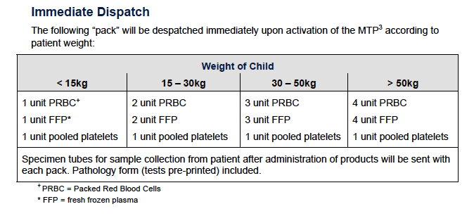
  • Massive Transfusion Protocols help and emphasise the need for not just the red stuff but good amounts of a fibrinogen source (locally that’s cryoprecipitate rather than fibrinogen complex concentrates, platelets and FFP. A quick Google search will find the guideline used at The Children’s Hospital at Westmead and the breakdown of what comes first…

Pack 1

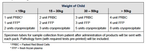
and what comes next…

Pack 2.jpeg

  • The number for pretty much all of the units (at least to start with) is 10 mL/kg. Quickly figuring out how much 10 mL/kg is for the patient in advance makes the calculations a lot quicker.
  • Of course the one different one is cryoprecipitate which is around 1 unit per 5 kg (up to 10 units).
  • Calcium replacement shouldn’t be underestimated as an ally (or even necessity). Perhaps me ending up mostly looking after kids just coincided with everyone getting interested in calcium, but I lean on this way more than I used to, particularly as the things that are supposed to help you clot go in.

Of course you’re not allowed to talk about trauma without mentioning tranexamic acid (TXA) because we’d all like to make sure there’s at least a little less bleeding if there’s a way we can influence it. So we want to get it there and get it here quickly.

The main question then is how much should we be giving?

Getting Bitten

The one guideline out there is the one from the Royal College of Paediatrics and Child Health. Back around 2012 they came up with a “pragmatic dosage” of 15 mg/kg as a loading dose then 2 mg/kg/hour.

I can sort of see why because there’s not a huge amount of evidence out there for ideal dosing in kids, particularly in trauma. What we end up with is evidence from other settings where traumatic damage is inflicted on tissues (i.e. big surgery).

If you go to any of those settings, like craniosynostosis surgery or scoliosis surgery or cardiac surgery, you’ll see a dizzying array of dosing regimes too. Loading doses of 10, 20, 30, 50 and 100 mg/kg with infusions any of 2, 5, 10 and 20 mg/kg/hr. This only makes figuring out what to do an awful lot harder.

So when they came up with that “pragmatic dosing” they went for a pretty cautious option. That’s partly because they’re not super sure about risks of thrombosis and there’s lots of concern about seizures with TXA loading. The theory goes that with higher doses you get higher levels of TXA in the CSF and that leads to inhibition of inhibitory glycine and GABA receptors (because they have those crucial lysine binding sites). It’s not everything but there’s at least some cohort research suggesting there’s not much association. In a retrospective study looking at craniosynostosis surgery with 1638 records examined the rate of seizures was the same across groups at around 0.6%.

The problem with that dosing option is there’s enough evidence to suggest that 15/2 is just not going to cut it. You might as well get a mosquito that bit a person who once had TXA and get them to sneeze on your patient. Bigger doses seem likely to work better.

A relatively recent paper in scoliosis surgery patients compared higher dose TXA with a lower dose. In this case the higher dose meant loading at 50 mg/kg then an infusion of 5 mg/kg/hr while low dose meant loading at 10 mg/kg then infusion at 1 mg/kg/hr.

So was there a difference? Well the lower dose crew lost an average of 968 mL and needed 0.9 units of red cells on average. The higher dose crew ended up losing about 695 mL and receiving 0.3 units of red cells on average. Unfortunately there was only 72 patients in the lower dose group and 44 in the higher dose group. So we’re left with not much.

There’s enough to suggest though that higher doses are probably required to actually influence the fibrinolytic pathway. A dose of 20-30 mg/kg to start with is much more like what I’d do (without exceeding 1 g) followed by an infusion of 10 mg/kg/hour.


The Next Bit

Look, don’t you think this has gone on long enough? Everyone did great, the surgeons operated really well and everyone got through a tough day pretty well and gave our imaginary patient the best shot possible.

There were of course other things we chatted about. Things like tricks for getting that IV access (if you remember the name Seldinger and that a 0.018” wire will fit up a 24 gauge cannula you’re in good shape). Then the challenges of spine immobilization and the role of options other than a hard cervical collar. Then of course the importance of considering the impact on ourselves when we look after these kids.

None of those deserve short change though so that can wait for some other time. Or maybe there’s an expert out there for that.


Notes:


The things on radiation risks in kids to look at would be this one:

Pearce MS, Salotti JA, Little MP, et al. Radiation exposure from CT scans in childhood and subsequent risk of leukaemia and brain tumours: a retrospective cohort study. The Lancet. 2012;380:499-505.

and this one:

Mathews JD, Forsythe AV, Brady Z, et al. Cancer risk in 680 000 people exposed to computed tomography scans in childhood or adolescence: data linkage study of 11 million Australians. BMJ. 2013;346:f2360.

Then of course there’s the bigger Royal College of Radiology Guidelines.

Oh, and the ATOMAC guidelines would be these ones:

Notrica DM, Eubanks III JW, Tuggle DW, et al. Nonoperative management of blunt liver and spleen injury in children: Evaluation of the ATOMAC guideline. using GRADE. J Trauma Acute Care Sure. 2015;79:683-93.

Here are those Brain Trauma Foundation TBI Guidelines. 

The kids TBI guidelines are here.

I can save you the Google search when it comes to that Massive Transfusion Protocol.

That RCPCH document about TXA in trauma is this one.

The thing in craniosynostosis surgery that covers seizure risk is this one:

Goobie SM, Cladis FP, Huang H, et al. Safety of antifibrinolytics in cranial vault reconstructive surgery: a report from the pediatric craniofacial collaborative group. Pediatr Anesth. 2017;27:271-81. 

The high-dose vs low-dose scoliosis study is this one:

Johnson DJ, Johnson CC, Goobie SM, et al. High-dose versus low-dose tranexamic acid to reduce transfusion requirements in pediatric scoliosis surgery. J Pediatr Orthop. 2017; 37:e552-e557.


Things that Go Up Kids’ Noses – THRIVE and Paeds

Nasal prongs seem pretty popular for lots of things these days. So how about their use in kids. There’s a couple of papers out there on its use in the paralysed patient and Dr Andrew Weatherall is here to splice them together. 

Isn’t it supposed to be the kids who stuff things up their noses? Have we just seen them do it so often we started wondering about the possibilities ourselves?

Let’s assume not. It’s more a case of people finally getting around to testing things out on kids when they’ve been running with them in adults for quite some time. This time it is THRIVE and that ever so desirable feature of endless maintenance of oxygen saturations while we get around to the ensnorkelling we’ve planned.

In principle that makes plenty of sense. The normal kid is more likely to rapidly desaturate than the normal adult. Physiology is pretty insistent on that. Plus we know people find paediatric intubation tricky so dropping the stress by avoiding the slide of the plethysmograph tone down that digital scale is probably a worthy pursuit.

So how about we look at two papers examining just this issue – does THRIVE employed in the little people stop those saturations from … not thriving??

travis-essinger-479636-unsplash
Fancy, nose-cramming air is what we’re dealing with really

Australian Angles

First up is this paper published by Humphreys et al, who work out of Brisbane. They did a small RCT on well kids with 24 in the control arm and 24 receiving 100% THRIVE. The kids fell between the ages of 0 and 10 years of age and are reported in the age groups 0-6 months, 6-24 months, 2-5 years and 6-10 years (with a total of 12 in each age range, meaning 6 in the controls and 6 in the THRIVE group within each age group – got it?)

The routine went something like *induction of anaesthesia* –> pre-oxygenation by doing that whole bag-mask ventilation bit –> the mask disappeared and THRIVE was added or nothing was added –> start the stopwatch.

You’ll note that, like the other paper we’ll mention, this is not about patients who are spontaneously ventilating. That’s a completely different thing.

In this group though the period of the saturations staying up was longer. Across the age groups the extension in apnoeic time was 86.8 seconds (0-6 months), 88.7 seconds (6-24 months), 129.5 seconds (2-5 yeas) and 169.2 seconds (6-10 years).

Right, lock it up. Everyone should have nasal prongs. All the time. It’d stop peanuts ending up there too.

Except there’s more pesky nuance in this paper. Like:

1. It’s not for pre-oxygenation

It’s worth noting that the preoxygenation here was all about face-mask ventilation with a good seal. They added THRIVE after that bit and started the clock. This is not entirely surprising because we know that nasal prongs compromise seals in adults and that’s only more likely with kids.

So if you were thinking that you should set up those nasal prongs from the before time zero, you need to think again. THRIVE for preoxygenation is not something tested here, and you shouldn’t assume it’d be better than good face-mask technique.

2. They didn’t test the duration that it worked for apnoea

All they said was it’s ‘more’. ‘Wait,’ you might say, ‘you mean they didn’t test the thing that was the point of study?’

Well not really because the cut-off was ‘twice the previously noted time to desaturation’. So they tested that they could reach the ‘double or nothing’ limit, but didn’t test the full extension. In the THRIVE groups the average saturation when they stopped the clock was 99.6%.

So I guess be reassured that it was likely to be really a heck of a lot of time.

3. Basic things were part of the procedure

For this study there was a lot of basics being done well. Throughout apnoeic oxygenation they weren’t doing things like airway instrumentation, suction, intubation or, I assume, anything much beyond chatting about the weekend and watching the clock. They did jaw thrust, a basic manoeuvre likely to optimise the impact of THRIVE. So maybe we should remember that all those things we are also interested in were not part of the picture.

And Now an Update from the Swiss

What if you didn’t make your cut-off ‘2 times the other cut-off we knew about’? How long could you go?

Well a Swiss crew with no interest in being neutral on the topic I guess have done a study comparing low flow nasal oxygen (0.2 L/kg/min) with THRIVE at either 100% or 30% FiO2 with 20 in each group. And they found … (wait for it…..) 100% THRIVE prolongs apnoea time.

OK there wasn’t much suspense there really.

Except again it was more subtle, and again cut-off matters. They had a cut-off to terminate on the basis of desaturation, but another at 10 minutes (as in ‘it’s 10 minutes and I’m bored let’s stop because those saturations are still great’) and the 3rd cut-off was if the transcutaneous CO2 hit 65 mmHg.

In the THRIVE 100% group no one desaturated, 4 hit 10 minutes and the other 16 had their nasal prongs ditched when they breached the CO2 target. This actually accords with the other paper where they also found that THRIVE doesn’t achieve ventilation and removal of CO2 in kids.

But at the end of this paper you still can’t say how long apnoea might be extended, at least when it comes to those saturations staying up.

Oh, and a couple of other points:

1. Pre-oxygenation was with face-mask ventilation. Again.

Again the nasal option had no role in the preoxygenation phase. They went with face-mask ventilation until the expired oxygen was 90% or above. Then they started the clock with the chosen nasal prong option going.

2. The other airway things done at the time were … none.

Yep. Once again this was just about the oxygen and the stopwatch. Nothing else was going on.

mihai-surdu-170005-unsplash
I mean this could be a visual metaphor for the need to appreciate their is still colour not just black and white when it comes to THRIVE or it could just be pretty, you choose. 

Let’s Think Clinically

So let’s imagine that we’ve actually got that paediatric patient in front of us. Maybe one who needs to get intubated before we get them out of wherever ‘in front of us’ is.

Let’s agree that maintaining oxygenation throughout is a good and noble goal. It’s not the only goal of course. We’d also like to make sure we make good choices around number of attempts, and for some patients (say the patient with intracranial pathology) we need to think about ventilation.

And we don’t have evidence that pre-oxygenation is aided by having THRIVE in place.

So assuming we’re going to do things standard to modern paediatric RSI like face-mask ventilation for a bit before we get going. There is at least a bit of  a question about whether THRIVE adds a huge amount.

What it undoubtedly adds is the confidence that saturations will stay up. That is something that lots of practitioners, particularly those not regularly intubating kids would find immensely reassuring.

There is a couple of caveats to keep in mind though.

There’s a risk to be aware of with THRIVE that those saturations staying interminably up might encourage tunnel vision on persisting with intubation when it’s not working out. It’s not too hard to imagine the scenario where the tube hasn’t passed straight down, but those saturations are OK so you persist a bit longer, and a bit longer, and now long enough that the airway is becoming traumatised and suddenly you’ve created a problem.

So this might be a cognitive challenge to have planned for in advance – how do you keep yourself to a limited number of attempts before re-evaluating and going to plan B (or C)? Do you make it a personal process or have others in the crew hold you to a maximum number of attempts or maximum duration of looking?

After all, THRIVE is going to get you to 10 minutes probably. But if you’re still conducting open negotiations with the glottic structures at 10 minutes, oxygenation is not the airway problem that should still be at the front of your mind. While you’re there, you might have to think about re-dosing anaesthetic agents too.

And the other key patient group is that one where intracranial pathology is an issue. Letting the CO2 rise for some patients is not a good plan because your TBI patient (as just one example) doesn’t need those cerebral vessels dilating and the intracranial pressure going up. For those patients, a step back to face-mask ventilation, or potentially placing a supraglottic airway,  to re-establish an ability to exchange CO2 is probably a better option.

So THRIVE might be great for some things. But whether it’s clinically better than an approach to the airway where really excellent pre-oxygenation is routine and good practices around face-mask ventilation are established seems like a line ball call.

I mean it’s still way better than a piece of Lego up the nose. But it remains an adjunct to the basic stuff, not a replacement.

Notes:

OK. That first paper is this one:

Humphreys S, Lee-Archer P, Reyne G, et al. Transnasal humidified rapid-insufflation ventilatory exchange (THRIVE) in children: a randomised controlled trial. BJA. 2017;118:232-8. 

The second one out of Switzerland is this one:

Riva T, Pedersen TH, Seiler S, et al. Transnasal humidified rapid insufflation ventilatory exchange (THRIVE) for oxygenation of children during apnoea: a prospective randomised controlled trial. BJA. 2018;120(3):592-99.

Did you want something on nasal prongs and seals? You could try this

Groombridge C, Chin CW, Hanrahan B, Holdgate A. Assessment of Common Preoxygenation Strategies Outside of the Operating Room Environment. 2016;23:342-6. 

or this

Hayes-Bradley C, Lewis A, Burns B, Miller M. Efficacy of Nasal Cannula Oxygen as a Preoxygenation Adjunct in Emergency Airway Management. Ann Emerg Med. 2016;68:174-80.

We’re always interested in other thoughts so feel free to drop a comment.

The Social Resuscitation

There are parts of the resuscitation with no algorithm. No protocol. How do we improve that part? What are the social resuscitation skills we need to work on? We’re very pleased to have Dr Ruth Parsell chip in with some thoughts. Ruth is a current ACEM Registrar working on the CareFlight Rapid Response Helicopter in Sydney. She joined the NSW Ambulance Service in 1998 and has worked in prehospital and hospital settings in varying roles since that time. 

The “social” resuscitation is a term I’ve been using for quite some time now. I apply it in dire situations. In both adults and children. But this is about the paediatric resuscitation and, specifically, cases where the prognosis is highly likely to be tragic. It is in these cases that I utilize this term because we are clearly treating more than just the patient when we resuscitate. I use the term because when I treat the child I am treating their family and all of the social connections that are linked to such a brief, precious life.

Experience We Don’t Always Want to Gain

The sad reality is that every paediatric resuscitation we do offers an opportunity to improve more than just our clinical skills. We all wish we didn’t see these cases but if they continue to occur then we will continue to do our best to serve the needs of both the patients and their families. What if we were able to improve the way we serve them? Which part of the resuscitation we call “futile” is the opposite of futile?

The best way to do both would be to have the “miracle” recovery. The “against all odds”, the “everything was against them”… the full recovery of a child who has had a terrible insult. The drowning, the fall, the pedestrian, the horse riding accident… all the terrible insults we see and all those mechanisms of injury that can potentially cause an early cardiac arrest or a moribund child.

Instantly we think of our algorithms, our protocols, our list of reversible causes and the sequence of steps we might take when we arrive at the scene. We hear the age, we think about weights, sizes, drug calculations. None of this should ever change and I’m not suggesting it should.

But what about when we hit that turning point?

It may have been an inkling early on. The thought that the mechanism is just too great, the injury just too severe, a poor response to even the most efficiently and expertly performed algorithm. It’s a moment where, sometimes even without verbalizing, the whole team is aware of the magnitude of the odds against this little one.

The Pause

What if in these cases we took a moment? Just a brief moment. When it comes to adult resuscitations I find we seem to automatically provide explanations to the family even while we are working. To explain that his heart is not beating and that we are working very hard to restart it; with a breathing tube, trying to stop the bleeding and with powerful medicines.

Perhaps it feels automatic because we just see more of those cases. We get to drill those algorithms more so there is a window that gives us space to look around.

So how do we provide this window in those paediatric prehospital jobs?

What if it was just a kiss before the transport? What if the family could have a little more from us? What if we suggested getting their daughter’s favourite teddy or blanket from the house? Just to fill their arms for the trip to hospital, to stop Mum’s hands from relentlessly wringing or something to give her tears a soft landing when they fall.

What do the books say?

The evidence for family presence during resuscitation has evolved over many years. Factors examined include the resuscitation team performance, stress levels amongst staff, clinical outcomes and psychological outcomes for family members. The evidence in paediatrics, including in some randomized control trials, demonstrates that there are improved measures of coping and positive emotional outcomes among families (1). These outcomes are achieved without impeding team performance.

There are many barriers to family presence in the pre-hospital arena. These scenes can be highly distressing, emotions are raw and the procedures required are time critical. Transport logistics can be a huge barrier too. It is rarely practical for a family member to travel with a child to hospital when they are critically unwell or in cardiac arrest. The confined environment of the back of an ambulance is usually congested and the potential unpredictability of a relative may compromise staff safety. The evidence regarding family presence is also more difficult to obtain.

However, there is some evidence regarding family presence during pre-hospital CPR in the adult literature and this also confirms positive results on psychological variables in family members without interfering with medical efforts, either clinically or with regards to health carer stress.(2)

When I have used the term “social” resuscitation in the past, I used it primarily in the dire situations I mentioned previously. Traumatic cardiac arrest in children fits this description, with a less than 5% neurologically intact survival rate (3).

I use this term in cases where I feel the resuscitation efforts are more a resuscitation for a family than the  patient. I use it in the context of transporting to an appropriate place, where I feel that the optimal ongoing social supports for family members can be best met. Somewhere where others can assist with tissues, quiet rooms and hushed explanations. Somewhere where others can understand the welled up look that we give them when we enter the bay.

Now I think that the social resuscitation needs to start earlier. A more conscious and deliberate effort. Maybe not every time. Not when you can feel yourself buckling under the cognitive load. Not when your emotions are so close to the surface you can’t get the words out. Not when the scene is like a powder keg and you might just be putting people at risk.

gabriele-diwald-201135

But in those paediatrics cases we need to make a conscious effort to find a window, even where the algorithm is crowding us a little more. That might be the part of the resuscitation that isn’t futile for those left behind.

Try the explanation. Try the kiss. Wait for that teddy. Just try it and let’s see if it improves our social resuscitations. It might even just improve things for all of us.


Notes and References:

  1. ANZCOR Guideline 10.6 Family Presence During Resuscitation, August 2016. 
  2. Jabre et al. Family Presence During Cardiopulmonary Resuscitation. NEJM. 2013;368:1008-18.
  3. Fallat et al. American Academy of Pediatrics. Policy Statement: Withholding or Termination of Resuscitation in Pediatric Out-of-Hospital Traumatic Cardiopulmonary Arrest. Pediatrics. 2014;133.  

That image is shared unchanged from the post by Gabrielle Diwald at unsplash.com under Creative Commons.


Just a Prick – Things that Might Just Work with Kids IVs

Putting a cannula in kids can be… well, an experience. Dr Andrew Weatherall has a collection of tips and tricks that might just be useful. 


Cannulas. Little people. Not always a match made in heaven. At the joint I work doing kids’ anaesthesia, we often note that they are the great leveller because it doesn’t matter how special you’re feeling, you’re just one lousy cannulation day away from feeling very, very mortal.

They are sort of essential for prehospital and retrieval work though. The thing is that we know that if you’re not working in a designated paediatrics job, the chances are that the little sprocket end of the market is by far the group you see the least. Which is not ideal for gaining and retaining skills.

So short of approaching random families in the street to see if the kids have always felt like their life was missing a cannula and would they like you to help with that (and that is a terrible start-up idea, don’t do that),  you have to make your best of the opportunities you have and draw on thoughts from other people.

So collected here are a bunch of things that help me get those little cannulas in. It’s not an exhaustive list of everything everyone has ever come up with of course. It’s just stuff that works in my hands that I’m sharing, partly in the hope that other clever people will chip in with suggestions in response. There must be some experts out there that we just need to poke enough to make them vomit up their wisdom.

I’m even going to leave out the “give them an anaesthetic and get them to sleep” one because it feels a little like cheating for this scenario.  And for the purposes of this post I’m not going into ultrasound stuff because that’s a whole extra thing. Let’s just put on record that if you’re cannulating for retrieval give it a strong thought.

So in a “not necessarily the most sensible order” kind of way, here’s how I’d think through that whole cannulation palaver:

1. What’s my aim here?

Knowing why you’re bothering with that cannula might seem like a dopey place to start but it sort of determines a bunch of decisions that follow. If you’re in a prehospital setting and you’re thinking of the cannula to get analgesia happening, do you have options you can start with first (intranasal or methoxyflurane etc) that will treat the clinical problem in the short-term and buy you time (plus help the kid, family and you) before getting to the cannula you might need long-term?

Are you adding one as a precaution for transfer? Is that the best choice for the patient and you? If it’s not time-critical do you have time for local anaesthetic options to do their thing?

Do you need the sort of urgent access that might befit an intraosseous option, then quick resuscitation and then an attempt at an IV once there are actually veins that have actual circulating volume in them to work with?

In this setting, it might well be that the IV is exactly what you need of course. But making that an explicit step in your thinking is a good thing. It makes you really prioritise the vital steps for management of the patient.

2. What’s my limit?

This flows from point 1. How many attempts would you consider before you try something new (like a different form of access, or asking someone else to have a go)? If it’s a cannula that must be done, your limits are going to be different than if you have nothing to start with. If you’re in a retrieval, rather than prehospital setting through there might be lots of clever people who can help (or who could do it while you do things that only you can do).

Setting some sort of soft limit where you will stop and reassess does stop you getting into the hole that comes with “I have to get this in” to the point where you forget the primary needs of the patient and it becomes mostly about pride. I’ve been there. A pride hole helps no one.

I don’t think you run the risk of mentally setting up with an assumption that your attempt will go wrong by having that limit either. It’s just about keeping whatever the primary goal of care (which is almost never the cannula itself, but what you can provide with the cannula) foremost in your mind.

Limit Vandys
Thinking and prep time might just save you a bit of time later

3. Super prep

Preparation is pretty much everything here.

The Patient

And whoever is helping them obviously but mainly the patient. If you’re with an awake patient, then telling them what you’re going to do and why is a pretty important place to start. The style that people employ for this can vary but one thing I’d be pretty firm on is that you can’t win by being dishonest. If it’s likely to hurt, don’t promise it won’t. If they’ll still feel pushing (like when you’ve used local anaesthetic cream), probably warn them. Let them know if you’re going to get someone to do the work of keeping a limb still. Explain steps as you go.

Positioning

If you can, choose to work in a position you find comfortable. Removing any degree of strain from your own posture just makes it easier to keep your later movements refined and precise. Not always possible, but working at the right height or even sitting down can make all the difference.

Look Everywhere

Way too often over my career I’ve gone to put a cannula in a spot because it seems convenient and later realised there was a much more accommodating vein somewhere else. There is something even more convenient than a vein that is close to where you’re standing. The vein that will actually help you out that’s all the way over there. Over that other side.

Check all 4 limbs, every time you can.

The Kit

Stuff for cleaning, stuff for doing, stuff for securing. Have it all ready to go (and that includes a back-up cannula ready in case you need to move on to another attempt). Once it’s in you want to be able to have it secured as quickly as possible. And once you’re under way you don’t want to be distracted by needing to reach for anything else.

A really good clean with an alcohol swab has an additional purpose. Sometimes it highlights a change in contour of the skin as the light picks it up and this reveals a vein. Sneaky and appropriate infection control.

4. The Actual Doing

Right. The pointy end. So to speak. Not so certain about this bit? Well these are all things I  do or have seen others do. Comprehensive? Probably not. For everyone? Maybe not but worth a think I reckon.

Choose your cannula

First up, examine that vein and decide which cannula you think will actually go in it. We all love a cannula big enough to rehydrate a woolly mammoth (and think how dried out those codgers would be now), but the truth of paediatric patients is that you don’t need a massive cannula to achieve good fluid loading. And you can definitely resuscitate more effectively with a smaller cannula in the vein than a bigger one in the subcutaneous tissues.

I’d even cope with a not-super-huge cannula in the cubital fossa if that’s what you need to get things rolling. At the hospital we regularly resuscitate kids without a huge cannula. It just needs a syringe and a 3 way tap (and you can actually do with most lines without a 3-way tap). Mostly it’s actually about paying attention and doing it, rather than letting it run.

Line it up

Absolutely the commonest thing I see trainees do when they are struggling is not actually lining up the cannula with the vein it is supposed to slide into. The entry point is somewhere near, but if you look at the barrel of the cannula, it doesn’t line up with the direction of the vein. Good luck with that.

Don’t focus so much on the entry point you forget the rest of the thing.

Make a hole

OK this one is probably more for the retrieval setting (and particularly for tiny ones) though I guess in principle as long as you have good sharps management you could maybe consider it for prehospital work (I’ve never done it there though). Not sure I’d try it in an awake child without some local numbing happen either.

After you choose your cannula, get a needle bigger than the gauge of the cannula. Make a hole in the skin at your entry point. Now when your smaller cannula passes through the hole you shouldn’t have the skin dragged in with it at all. You should lose all resistance at that level actually. Do it right and pretty often you’ll feel the end of the cannula pop into the vein before any visual clue like a flashback tells you that you’ve made it.

Note that having gauze handy for any small amount of blood ooze that would obscure the entry point is helpful here.

Petras Gagilas
Look, not so big you can see light through it, but something.

The saline trick

I think this only works with non-safety cannulae. Basically you fill the hub with saline and when you hit a small vein you’ll see a super quick flashback (even just starting with a quick change in the light in the saline). This one’s particularly useful for getting early warning in tiny veins to avoid going straight through.

Short and sharp

You probably understand that you need to come really flat to the vein with your angle of approach (by all means be at a more acute angle to get through the skin, but approaching the vein should be pretty flat).

The other key bit though is short, sharp movements forward followed by a pause. I tend to find slow advancing just doesn’t do the job in little people’s veins. It’s like the slow distortion of the tissues encourages them to roll out of the way (you can even see it on ultrasound). A sharp move forward, then a pause, then repeat just seems to work better.

The Roll

You get the flashback. Victory! Except you still need to advance and you’re worried it’s a bit small that there vein. So do a really small advance. Then rotate the whole cannula (as in the needle bit as well) 180 degrees. The leading edge of the needle is now closest to the skin, and the pointy bit isn’t going to go ahead and spear the back wall. Advance a little more. Now feed off the cannula.

The Twist

This trick is more well known. Once you think you’re in that vein, twist the cannula off into the vein. In bigger kids it’s probably no help but in smaller veins it does seem to sometimes help get it not to catch up on the wall of the vessel.

Wired for Not Sound

This one is not really a prehospital thing but if in a retrieval-type situation you could consider this one. Have a think about getting familiar with wires for Seldinger options. There are manufacturers out there making short wires that will feed down a 24 gauge cannula. Arrow make one that is 0.018 inches (diameter) and Cook make one even smaller (at 0.015″). When you have one of those cannulae you really want but after you feed it off it’s all gloom, a wire can rescue you.

The technique (with appropriate cleanliness and wire precautions to ensure you don’t lose it in the vein all in place) is to gently start pulling back just the cannula until you have blood freely flowing back. If you gently advance the wire up the cannula at this point it will sometimes find its way perfectly up the vein. If so, you now have an introducer to place a cannula (maybe even one larger than the one used for access).

Not a technique to try in anger for the first time without someone who has done it nearby I’d say.

It’s also worth noting that not all wire/cannula relationships are without challenges. For whatever reason a Surflo 24 gauge cannula will absolutely not allow a 0.018 inch wire through. A 24 gauge Insyte? Well they were made for each other. Go figure.

4. The Strapping

Well that’s a completely different post. I only wish someone had good tips for things like that (like say, here).

For kids cannulas there are a lot of techniques out there and lots of strong opinions about tape. My main thoughts would be:

  • The tape has to be in contact with the actual thing it is supposed to hold. Sometimes I see people holding tapes tight as they put it across the cannula, thereby guaranteeing the tape only contacts the top surface and is then stretched onto the skin. Form the tape closely to the cannula itself. Squeeze it right on there to get maximum tape-to-cannula contact. Then lay it across the skin (no stretching) and put some pressure on it to get adherence happening.
  • Really think hard about things like boards. If they are not adding security for that cannula, you can almost guarantee they are adding annoyance for the patient.


So there’s a start. I bet people have more I’ve forgotten or don’t even know about.

You might just find some of these tips help though. And if that’s the case you will hopefully end up not being the big prick finding it a bit of a prick to get a little prick done for a little kid.

Little kid. What did you think I was going to say?

Notes:

I am not kidding about hoping people will have better tips. That’s what the comments bit is for. Go nuts. Or share the post and see if someone else has one.

Also, if you like the posts here remember you can sign up to get emails whenever they drop. It should be here on the page somewhere.

The images here were from vandys (the speed limit one) and Petras Gagilas (the tunnel thing) and used unchanged from their spot on flickr under Creative Commons.


Old School/New School – Updating Classic RSI

Respect for the classics doesn’t mean being stuck with them. Here’s a refresher on why you might not want to do RSI like they used to by Dr Andrew Weatherall. This one is a cross post picked up from the paeds anaesthesia site he chips in on, www.songsorstories.com 

Everything in medicine needs the occasional reboot. I mean not as often as Hollywood thinks we need to reinvogorate a superhero franchise but at least every now and then. Sometime that’s because we learn new things (cross reference here). Sometimes it’s because our perception of what is the biggest risk changes (more on that in a second). And sometimes we suddenly realise that the original reason something became fixed practice might not have been a thing in the first place.

Which brings us to RSI, a classic so many of us have grown up with.

What is this thing?

The story of RSI starts with excellent intentions (and for this version of events I’m leaning heavily on this review by the excellent Thomas Engelhardt). In this case the idea was to come up with a safer way to get the snorkel in the all important windpipe as quickly as possible to try and minimise the risk of things that should stay nestled in the gastrointestinal tract might find their way to the lungs.

And you can understand why. Serious aspiration can, sometimes, be deadly. The first piece of the puzzle was written up by Morton and Wylie way back in 1951 who described where with the patient sitting up the anaesthetist would give intravenous barbiturate then muscle relaxant and rapidly intubate them. A rapid sequence of induction and intubation. So really it’s RSII.

8 years later a description emerged of a thiopental/relaxant/40-degree head-up tilt foot-down tilt. It wasn’t for another 2 years that cricoid pressure popped up (thanks Sellick) although interestingly it included not just a bit of pre-oxygenation but also some bag-mask ventilation prior to putting the tube in.

It was another 2 years before the other classic bit of RSII became popular, with an exhortation to avoid bag-masking because of the perceived risk for gastric insufflation and hence regurgitation.

A classic technique derived from a series of “what abouts” and “I reckons”. I mean, you wouldn’t read about it. Except you just did.

That’s not to say that medicine doesn’t have space for a bit of logical derivation of good ways forward. It might just suggest that the whole approach is open to a refresh.

Re-evaluating the Likely

If the technique was designed to prevent aspiration, maybe we should start with looking at how likely this event is in a setting a bit more modern than 1951. In 1999 the epic writing team of Warner, Warner, Warner, Warner and Warner looked at 56138 patients under 18 having procedures (elective or emergency) over 12 years to see just how big this problem was. This covered 63180 procedures.

The time frame for defining aspiration was entry into the operating room until 2 hours post-anaesthetic. To score the label there had to be direct identification of bilious secretions or particulate matter in the tracheobronchial tree or new X-ray findings after an episode of regurgitation.  A total of 24 patients met the criteria.

11 of those were emergency cases so the rate in that group was 1 in 373 compared to 1 in 4544 in the elective cases. 21 of the 24 were around induction. 15 of the 24 had no symptoms develop despite the aspiration. 5 of the other 9 did need respiratory support of some kind and 3 of them needed ventilation for more than 48 hours. Well the paper says that but actually describes ventilation for 18 days, 14 days and 33 days in those cases.

And there’s the rub. It’s really very impressively rare. But then when it goes bad, the downside can be very, very down.

So fine, let’s prevent the bad thing. We’d better get on with the classic old RSII, right?

Remembering the Even More Likely

The problem with being so rigorously focussed on avoiding pulmonary aspiration that you do things like not help the patient breathe, is there are other basic functions that don’t get looked after so well. Like oxygenating.

Gencorelli et al looked at episodes of desaturation during RSI while describing the classic drugs/cricoid/no ventilation technique. Across 1070 children included they reported a 3.6% rate of desaturation to 89% or below (1.7% of the patients being in the under 80% group). Not surprisingly the under 2s were more likely to have a desaturation.

These rates are low of course and certainly lower than in some other areas of practice. Reports from emergency departments have indicated desaturation rates anywhere from 14% to 33% (with the latter reporting rates of desaturation of up to 59% in the under 2s).

So amongst the various things we’re trying to do to prevent the 1 in 400+ event are we at risk of failing on another key thing. You know? The oxygen provision thing.

What’s the alternative?

Neuhaus and team subsequently described very well their approach to RSII, which they badged as cRSII (where the “c” is for “controlled” not some other “c” word like “cheese” which wouldn’t make sense anyway but would be a good reminder that cheese is great).

They key features for them (putting to the side “lots of preparation”):

  • 20 degrees of head up (though they say only for the over 2s)
  • Suction any NG in situ.
  • Give the drugs.
  • Avoid cricoid pressure (with a few exceptions).
  • Provide gentle facemark ventilation with peak pressures of 12cmH2O.
  • Neuromuscular monitoring to ensure the muscle relaxant has really, really worked.

This last point makes a heap of sense as active regurgitation is a problem created by airway instrumentation when you don’t have adequate anaesthesia and paralysis.

cRSII
It’s a big list.

Talk is cheap though, what were their results?

They report on 1001 patients They had a moderate hypoxaemia (89-80%) rate of 0.5% and a severe hypoxaemia (< 80%) rate of 0.3% and the 8 patients this represents had a median age of 0.8 years. They had 1 patient with regurgitation but no evidence of aspiration.

That’s pretty impressive.

Putting it Together

So if we accept that we should really try and optimise oxygenation, and that the risk of this is higher than the risk of aspiration then we have to accept that modifications to that original technique are reasonable. What are a few steps for practically putting it together?

1. Assess that risk of a full stomach

It might well be that we’re going to avoid cricoid most times, but there are still a few situations where that risk of aspiration is probably higher. In the Neuhaus paper they suggested achalasia, Zenker diverticulum or post-colonic interposition patients (done for oesophageal replacement) always need cricoid.

It certainly seems worth having heightened concerns in the patient with significant increases in intra-abdominal pressure.

2. Everyone sits up

Why wouldn’t you have a bit of head up? It makes sense if you’re avoiding passive regurgitation and is a good position for pre-oxygenation, facemark ventilation and intubation. I’m not quite sure why some authors have suggested the under 2s shouldn’t be head up. This is a routine option.

3. Have that suction handy

Goes without saying maybe, but I’m saying it.

4. Pre-oxygenation, but not with distress

Yes you want to pre-oxygenate. And most times you can talk kids through that and get a full 3 minutes in. Some kids will only get more distressed with oxygenation though, and insisting on pre-oxygenation only guarantees distress. Given that you’re going to apply gentle face-mask ventilation, it’s rare you need to go to the wall on this one.

And while I’m there what about apnoeic oxygenation? Well, as discussed in this post, the evidence that’s available in kids isn’t so persuasive as to suggest it should be routine. The stuff that has been done showing extended apnoeic time actually followed effective pre-oxygenation with face-mask ventilation. So as we’re going to put that tube in quickly after the same sort of effective face-mask ventilation, extending apnoeic time for minutes seems not that clinically relevant.

5. Cricoid yes or cricoid no?

Again this is a judgment call. I know plenty of anaesthetists who still prefer to start with it but with a low threshold to remove it. I’m more likely to mostly err on the side of not using it, except for those high risk of aspiration patients.

If you are going to use it, it is worth noting that, particularly in infants, the trachea is quite often more prone to distortion by cricoid pressure than you realise. Doing flexible bronchoscopy work you’re sometimes asked to manipulate the airway and I’ve seen the whole trachea get substantially compressed and distorted by seemingly innocuous manipulation. Distort it enough and you can increase the resistance to air going in and out enough to make it easier to get down to that stomach.

In addition, as covered very nicely in this review, cricoid relies on the alignment of trachea and oesophagus and the evidence is that in kids < 8 years old 45% had displacement of the oesophagus so you’d be unlikely to get compression of the oesophagus even with perfectly delivered cricoid (at least on the CT scanning mentioned).

So for the very high risk ones I’d tend to start with it (well start with it once I’m sure the kids won’t react to it going on), but that leaves almost everyone where I would’t be too concerned. And if it is on, I’d be quick to take it off if it was impeding either view or tube passage.

OLYMPUS DIGITAL CAMERA
Maybe I included this picture of an echidna because they have a reputation for being good at waiting and not because it’s a prickly situation.

6. Wait

We’re going to take our time with face-mask ventilation and maintain oxygenation. So where’s the extreme rush getting the tube in? Being too obsessed with that step, even though you’re achieving oxygenation, is a way to end up instrumenting the airway while the patient is only lightly anaesthetised or inadequately provided with paralysis. What was that thing we’re preventing again? The regurgitation thing that’s worse if we get going while the kid is lightly anaesthetised? Oh, right. Slow down.

The description suggests using a nerve monitor. I can’t say this is routine myself, but once the muscle relaxant is onboard I do publicly note for the team I’m working with how long we’ll be waiting on the clock before we start trying to intubate. (“The clock says 09:30 now. Once it ticks over to 09:32, we’ll start with the intubation.”)

I then remind everyone that this will take an unnervingly boring period of time and they might want to come up with a good joke to fill the time.

7. Ventilating

Yes, this is a thing that’s necessary because kids desaturate quickly. Particularly the younger ones. Achieving gentle face-mask ventilation relies on really good technique with the bag in hand. Plus it’s very therapeutic to gently squeeze that bag.

7. What about parents?

This one also needs an assessment of what might help and what won’t. For lower risk kids, as a paediatric anaesthetist doing it regularly, I’d be comfortable having them along. But if it was the sort of case that was likely to be difficult, or if I was back at the training junior doctor stage, there’d be no dilemma for me. I’d tell the parents that they wouldn’t be coming in. Having them alone to help their child relax (not always a guaranteed result of having parents in) has some advantages. But the prime job is safe management of the peri-induction period. And that might mean less people around.


So those are the simple things that have shifted over the course of my time in the big wide medical world. It’s a realignment of the priorities in a way that makes the ‘R’ in ‘RSII’ look smaller and smaller so that the oxygenation is placed at the top of the tree.

Put together though it’s a reboot worth endorsing. I mean the 60s just weren’t that great, surely?


Notes:

How many bits that are really important aren’t covered here? There must be some. So leave a comment. We’ll all learn.

And if you like the post and other things around the joint, maybe throw your email in the relevant spot so you’ll get an email each time a new post pops up.

This post is a cross-post from another site that this Weatherall bloke works on called Songs or Stories. It’s about paediatric anaesthesia.

That echidna pic came from flickr’s Creative Commons area and is unchanged from Duncan McCaskills’s post.

Now to the literature, because going to the direct papers is always rewarding.

That review by Engelhardt where he makes it clear what he thinks is this one:

Engelhardt T. Rapid sequence induction has no use in pediatric anesthesia. Pediatr Anesth. 2015;25:5-8. 

The paper by the anaesthetic equivalent of the Brady Bunch or something I assume is this one:

Warner MA, Warner ME, Warner DO, Warner LO, Warner JE. Perioperative Pulmonary Aspiration in Infants and Children. Anesthesiol. 1999;90:66-71. 

The benchmarking study is this one:

Gencorelli FJ, Fields RG, Litman RS. Complications during rapid sequence induction of general anesthesia in children: a benchmark study. Pediatr Anesth. 2010;20:421-4. 

The emergency department studies mentioned in passing for their demonstration of high rates of desaturation are these ones:

Long E, Sabato S, Baby FE. Endotracheal intubation in the pediatric emergency department. Pediatr Anesth. 2014;24:1204-11.

Rinderknecht AS, Mittiga MR, Meinzen-Derr J, Geis GL. Kerrey BT. Factors Associated with Oxyhemoglobin Desaturation During Rapid Sequence Intubation in a Pediatric Emergency Department: Findings from Multivariable Analyses of Video Review Data. Academic Emergency Medicine. 2014;22:431-440. 

That paper looking at controlled techniques in kids is this one:

Neuhaus D, Schmitz A, Gerber A, Weiss M. Controlled rapid sequence induction and intubation – an analysis of 1001 children. Pediatr Anesth. 2013;23:734-740.

And that other review is this one:

Newton R, Hack H. Place of rapid sequence induction in paediatric anaesthesia. BJA Educ. 2016;16:120-3.


The Dangerous Little Details

A new bit of research is out looking at paediatric intubation in the prehospital and retrieval setting. Picking it up and turning it this way, that way and all around, here’s Dr Andrew Weatherall. 

Advanced prehospital practitioners that I’ve met have some pretty common traits. They are pretty comfortable around things that other people might find chaotic. They often have pretty strong opinions on food and coffee. Not necessarily even on good food either. I’ve been given connoisseur-level education on various take away options. Most importantly, they are appropriately bananas about doing a good job for their patients.

That extends to paediatric patients which is obviously excellent. Except we tend not to do our most excellent work when it comes to kids. The reasons for that could fill many a blog post (and maybe we’ll get back to that another time) but kids tend to get less pain relief when faced with similarly painful situations, less interventions even when they’re indicated and we tend to do those procedural things less well.

In 2011 Bankole et al. compared interventions in kids (defined as < 12 years old) and adults with a head injury and a GCS < 15 in New Jersey (there was 102 patients in the kids group matched to 99 adults with equivalent injuries).  69.2% of the kids had some sort of problem with intubation. That was across failed intubation (29.03% vs 2.27% in adults), tube dislodgement (16.12% vs 2.27%), wrong-sized tube (7.45% vs 0%) and multiple attempts (as in over 3 tries) at intubation (6.45% vs 2.27%). A peripheral IV was there in 85.9% of adults but only 65.7% of kids.

In a paper that also commented on relative intubation rates in advanced EMS vs general EMS in the Netherlands, Gerritse et al also commented on analgesia. In their study 77% of kids who really needed some form of analgesia actually received nothing from the general EMS. No kid under the age of 4 received any form of analgesia from the EMS. Not one.

I’m not quoting those papers to say anything other than good practitioners (I have a predisposition to think most of those working at any level of EMS are people trying to do the best job their system and training allow) find kids extra difficult. This patient group provides an additional challenge on top of the storm you already deal with the scene. Like someone started blasting fairy floss into your eyes in the middle of that storm. OK I’m not sure that was the greatest analogy but it’s happened now so maybe we can just agree to move on while also remembering that when you’re a kid fairy floss is pretty great. Mmmm, fairy floss.

Enter the Swiss, purveyors of good chocolate and cheese with holes, with some interesting work that sheds a little extra light on things that even the most advanced practitioners find challenging about little people and airway management.

Let’s Stop and Check the Scenery

Not the mountains or lakes or Large Hadron Collider scenery, the other scenery.

Appearing in SJTREM, the paper comes from a  look at their database between June 2010 and December 2013. Across their 12 bases and one affiliate base they do around 11000 prehospital or interhospital missions per year with their paramedic-doctor teams. I should point out that these advanced teams really have had good training in airway management and specific paeds time. The study looks at any kid under the age of 17 requiring any airway manipulation (not just intubation or supraglottic airway or tracheostomy but bag-mask ventilation as well).

From their pool of 4505 paediatric patients over the 3.5ish years (which if they’re doing around 11000 jobs per year should be around 11-12% of their total workload) the ended up with 425 kids requiring some sort of airway care (9.4% of the paediatric group). A little over half (225) were prehospital cases. From here on in when we talk about intubation it’ll be about prehospital missions because those moving between buildings were already intubated and ventilated.

So what did these top operators find?

Actually It’s Not About the View

In the 215 patients for whom an attempt at endotracheal intubation was attempted, first-pass success was 95.3%. Now, if you’ve dropped by this blog before you might recall Dr Alan Garner discussing whether this is the most important measure. I think that’s a great post, but I don’t think it is meant to be interpreted as “first pass intubation tells us nothing” (Alan can always correct me).

What this number does say is that the challenges in kids aren’t necessarily about getting a view of the cords that is enough to achieve intubation. Only 10 patients (4.7%) were described as inflicting a difficult airway management scenario on the team. 98.6% eventually ended up with a support snorkel in their trachea.

There were 2 children who could not be intubated and ended up oxygenating very nicely with the aid of a supraglottic airway, while one patient with a known “airway issues syndrome” (Goldenhar’s syndrome) couldn’t be either intubated or ventilated but was already at the end of a prolonged arrest situation.

So for advanced EMS providers, maybe it’s not the getting a view/passing the tube part of the procedure that is really at issue. In our own research that touched on this, the intubation success rate was 98.7% of the paediatric patients were successfully intubated while one patient was managed with a laryngeal mask in the prehospital phase.

This fits with the overall truth of paediatric airways: unanticipated difficult laryngoscopy is less common in kids than adults.

So Where’s the Problem?

The problems with paeds airway intervention here are about the details. You may have noticed that people who do subspecialty work in paeds can be a little bit fanatical about details. There’s a reason for this. A smaller airway is less forgiving of the tube that is the wrong size, be it too big or too small. An endotracheal tube that is 1 cm too far in on your 1 year old is proportionally a lot closer to the carina than when the same situation applies to an adult. Add a little flexion or extension and that whole tube can end up visiting new pockets of the bronchial tree.

This is the part that is really well covered in the Swiss study. In the 82.7% where intubation was noted, 82.5% got an adequately sized tube. It was too shrunken to be appropriate in 2.9% and too gargantuan in 14.6% (in the under 1s that rose to 57.5%). Rates were higher if that tube was placed during a CPR scenario.

The depth? Well, if you went off the formulae often mentioned in dispatches, most insertions were deeper than that. And while I can’t seem to find the bit in the results that clarifies this statement, the authors say in the discussion that “Only the placement of the depth marking of the correct Microcuff ET tube … for age between the vocal cords was accurate for all paediatric patients …” (Not familiar with the markings? You could look at an earlier post on this site, here.)


Details, Details

I think this is the key message of this study. Lots of things might make you sweat about paediatric airways. I suspect that for most practitioners it is the view and “plastic through the cords” components that cause the stress.

That bit is important, of course, and everyone wants to do that bit well. This study supports the argument that advanced practitioners already do that bit really well. Perhaps in thinking keenly about that bit it’s attention to some details, the sort of details that kids are pretty unforgiving about, that gets in the way of safer paeds airway management.

Stavros Markopoulos
Look at this butterfly. Gets fuzzy on the last few details of the right wing and can’t even butterfly properly.

Things to Take Away

Any research only reveals a very particular part of a story. There are questions left unanswered or things that don’t quite apply to your practice. That doesn’t mean we can’t use those results to reflect on things we do when we deliver our variant of advanced care.

So I’d say there are a few key things suggested by this study:

  • If you’ve trained in paediatric airway management, chances are the intubation itself (at least the getting a view and passing the tube bit) will go well.
  • Really well trained people still find the details challenging. The wrong tube size and the wrong depth of insertion matter in these patients.
  • It might be time to review whether those old formulae are the best option.
  • Knowing your equipment (like where the line on the tube goes) is pretty worthwhile.
  • The tube through the cords isn’t where attention to detail stops. That’s not the moment to ease up.

So we can all get out there, push through the fairy floss, be confident that we’ll get those endotracheal tubes in and start remembering the little details that will produce perfection.

No more fuzzy butterflies.

Notes:

Of course it’s not the fault of the butterfly it’s right wing looks fuzzy. It’s the photographer. Well, actually it’s an amazing photo where the wing is a tiny bit in a different alignment. It’s from flickr Creative Commons via Stavros Markopoulos and is  unaltered.

The source paper link is right here and it’s open access:

Schmidt AR, Ulrich L, Seifert B, Albrecht R ,Spahn DR, Stein P. Ease and difficulty of pre-hospital airway management in 425 paediatric patients treated by a helicopter emergency medical service: a retrospective analysis. Stand J Trauma Resusc Emerg Med. 2016; 24:22. 

I also mentioned a paper we put out there:

Barker CL, Weatherall AD. Prehospital paediatric emergencies treated by an Australian helicopter emergency medical service. 2014; 21:130-5. 

Then there’s the Bankole et al. paper:

Bankole S, Asuncion A, Ross S, et al. First responder performance in pediatric trauma: A comparison with an adult cohort. Pediatr Crit Care Med. 2011;12:e166-70. 

And finally the Gerritse et al. paper which is also open access:

Gerritse BM, Schalkwijk A, Pelzer BJ, Scheffer GJ, Draaisma JM. Advanced medical life support procedures in vitally compromised children by a helicopter emergency medical service. BMC Emerg Med. 2010;10:6.

Addit: After a really helpful comment from Paramedidad the line “In their study 77% of kids who really needed some form of analgesia.” was fixed to read “In their study 77% of kids who really needed some form of analgesia actually received nothing from the general EMS.” 


Getting Things Straight

Lots of beliefs are hard to shake. Andrew Weatherall covers one from the paediatric airway – the holy status of the straight blade.

As I’ve mentioned before, paediatric airway management is full of mythological beasts. Some of that is about anatomy stuff and the like. Some is about equipment. Plenty is about technique. Sometimes it’s about technique and equipment together. Bliss.

So this is where I wade into another topic in paeds airways:

Straight blades are overrated and you should throw them away.

Marc Zimmer Dog Unicorn Dog
It is a time for mythical beasts. Like the fabled unicorn dog but less cool.

Do we need big bins?

Well, actually no. Stop the indignant letter writing. When I say they’re overrated I don’t mean they have no value. They have a role like most items of equipment that are still in use after nearly 100 years probably still have a role.

What I do mean is that straight blades are treated with a reverence in paediatric airway management that is unwarranted, while curved blades like the Macintosh seem to be described as “bigger people’s airway devices”. Trainees could easily go through their whole training period thinking that you must always use straight blades for patients who understand what the hell Pokemon are all about.

That just isn’t true. People who swear by straight blades will point to the more anterior epiglottis and the angle of the cords to argue the case for their chosen device just as convincingly as those who like a curved blade point out that they get more working space in the mouth and a familiar blade and both will be sort of right.

It might be useful to dive into this a little more. So let’s work through a paper from 2014 that specifically looked at the straight vs curved blade question. Partly because it gives an appropriate ‘meh’ when trying to split the two options but also because it highlights how myths can dominate our perception of the original work.


Welcoming the Contenders

The paper here appeared in Pediatric Anesthesia in 2014 (I touched on this in the other post). The authors set out with a useful question: is there a difference between Miller and Macintosh blades when it comes to ease of obtaining a view and success of intubation in the 1-24 month age range?

They looked at well kids having elective surgery under anaesthesia where muscle relaxation was also used. They included 120 kids and each kid had laryngoscopy with one device then the other.

The results are a case of a big old shrug, which is sort of OK. Easy laryngoscopy was noted in pretty much the same percentages. First pass success pretty much the same. The rates of one being better for the view than the other were pretty much the same. When it was difficult with one view the rate of switching to the other and finding it was easier was about the same regardless of whether you had started with the Macintosh or Miller.

So the two blades that stepped into the ring step out with no knock out punch thrown. There are a number of other interesting points when you look in more detail though and a few comments I’d make in passing.

  1. The epiglottis isn’t the endpoint

I have this impression that trainees get really obsessed needing to pick up and control the epiglottis with a straight blade. In this paper the routine use of the straight blade was to place the tip in the vallecula. In only 2 of the 60 uses of the Miller blade did they pick up the epiglottis.

Why do people get so antsy about picking up the epiglottis? It was only ever described as one of the options to obtain the view, not the only option. Those early designers never forgot the aim: to obtain a view to let you instrument the trachea.

Here’s Miller from the paper where he described his blade:

“The epiglottis is visualized and raised slightly to exposure the cords or, if the operator desires, the tip of the blade maybe placed in front of the epiglottis and raised sufficiently to visualize the cords after the method of Macintosh.”

In fact Macintosh described the straight blade being used in this manner when he reported on his own design, singling out Dr Margaret Hawksley as an exponent of this technique. The authors of this recent paper further point out that it’s a lot more stimulating to pick up the epiglottis. That’s worth at least a thought.

Miller wasn’t precious about how you get the view. The idea that picking up the epiglottis is the only technique just got repeated enough that no one remembers to question it. The epiglottis isn’t the main game. The view is the thing.

  1. Make sure of your basic technique

One of the other interesting features here is that there are some elements of the intubation technique that seem like they could do with a review. An example: the Miller blade was advanced centrally along the tongue. This is a technique taught by heaps of people and I think Miller probably would have strong feelings about that. Again let’s go back to the paper:

“The blade is inserted in the right side of the mouth, pushing the tongue to the left.”

One of the bigger challenges in getting a view in paediatric patients is getting the tongue out of the way. This is particularly for straight blades which tend to have less of a flange to do some of the work for you. I’m not the only one who thinks so, either:

“On passing the instrument into the mouth the tongue should be manipulated to the left side, away from the slot; otherwise the organ may roll into the barrel and completely obstruct the view.”

That was Magill. In 1930. Now Magill might have been describing the use of a speculum but the principle is the same. The tongue is only likely to make your view worse (and given that straight blades pose an additional challenge in having not as much space proximally to work in, that really matters).

Magill went on to point out that if you struggled at all you could move the proximal end of our instrument further to the right corner of the mouth – that’s also known as the paraglossal view and turns out to be pretty much the best way to go.

In other basic technique points the authors of this recent paper mention that laryngoscopy was done with the head in a neutral position. This doesn’t seem like optimal head positioning for use of either blade, and that’s another point worth keeping in mind.

  1. It’s useful to know what the intended use was with your instrument of choice

In this paper and in the comparison with the Cardiff blade they refer to as an example of other “blade vs blade” papers, a comment is made that when you introduce an endotracheal tube centrally you can compromise the view and that it’s not great for introducing your tube via any central channel.

Miller, again:

“The scope is used for visualizing the cords only. One should work outside the blade to insert the tube. The only criticism of the instrument has been that it is too small through which to work. It was not designed to be used as a guide for the catheter.” (That’s the author’s work with the italics, not an edit from me.)

In fact Miller searched for a new design because he felt small laryngoscope blades on the market were too big. It’s designed to be small.

So yes, you need to use a technique where you bring the tube in from the side. That was always the point.

  1. Should we stop talking about external laryngeal manipulation like it’s an extra?

This is really a bit of open musing on my part. This was done in over 50% of the patients in both groups and generally helped when it was used. I can see how the precision of description goes up when we include these details but it strikes me as so much a part of every intubation (as this external pressure means less work for the laryngoscope itself) that I wonder how much it adds to our appreciation of clinical use. That’s one for the comments section I guess.

Messages for the Prehospital or Retrieval Type

After sifting though all of that, what are the take home messages? Well, here are some from me that might need additions from others:

  1. Know what you do and why you do it

Those picking up a laryngoscope for the little people need to have thought through what they will use and why. If I’m offered a personal preference it is to use a curved blade for everyone. Even as a paeds anaesthetist I’ve just used a curved blade more. It’s better designed to control that pesky tongue. You get a huge working space within the mouth and with external laryngeal manipulation (which I’d call standard) you can pretty much always bring the airway into view, even in the slightly anterior larynx.

I haven’t seen a study that would confirm this hunch, but I wonder if one of the problems some prehospital clinicians have with paeds intubation is they pick up a laryngoscope they didn’t really learn, very rarely use or rehearse with and don’t really understand. You need to focus your technique slightly differently with a straight blade. Add the stress of the situation and is it any wonder the job becomes harder?

I’d back the occasional proceduralist as more likely to intuitively understand the anatomy using the same sort of blade they always use. I doubt it’s a study that would be easy to set up in anything but mannequins though.

  1. Know the different options

That preference for people using what they know doesn’t mean you shouldn’t learn both. This study did highlight that some kids just have a better view with a particular blade. You can’t quite get as good a view with one option, switch to the next and all of a sudden it’s easier. Again though if you’re reaching for that other option use it right.

  1. Make your technique appropriate for the 1% and the 99% will be fine

This is more of a general point. Laryngoscopy in infants is easy the vast majority of the time. So if you don’t bother controlling the tongue you’ll probably get by most times. It’s the 1% where your routine practice of not getting the tongue out of your view, or being able to aim for either the vallecula or epiglottis, or positioning the head right will start to bite.

If you always do everything to maximise your view, you’ve already got a good technique for the 1%. It’s best not to need to review your technique once the blade is in and you’ve figured out this is the tough one you’ve been dreading.

So after all that, maybe paediatric airway instrumentation comes down to a really simple refrain: the tool in the hand matters less than the tool holding it.


Notes:

That image comes from Marc Zimmer on flickr under Creative Commons and is unaltered.

Here’s the paper mentioned again:

Varghese E, Kundu R. Does the Miller blade truly provide a better laryngoscopic view and intubating conditions than the Macintosh blade in small children? Pediatric Anesthesia 2014;24:825-9. 

And the others …

A review on the popularity of the Macintosh blade:

Scott J, Baker PA. How did the Macintosh laryngoscope become so popular? Pediatric Anesthesia 2009; 19 (Suppl 1):24-9. 

Miller RA. A new laryngoscope for intubation of infants. Anaesthesiology 1946;7:205-6.

Macintosh RR. A new laryngoscope. Lancet 1943;1:205.

Magill IW. Technique in endotracheal anaesthesia. British Medical Journal 1930;2:817-20.

and the Cardiff paper:

Jones RM, Jones PL, Gildersleve CD, et al. The Cardiff paediatric laryngoscope blade: a comparison with the Miller size 1 and Macintosh size 2 laryngoscope blades. Anaesthesia, 2004;59:1016-9.


Teaching an Old Dog New Tricks – Or A Visit To An Alien Planet

One of the excellent things about retrieval work is the opportunity a clinician is presented with to try new things. Dr Alan Garner reflects on his recent experiences trying out a very particular branch of retrieval medicine – neonates and paediatrics. 

I have recently had the opportunity to do some work with NETS in NSW due to some staffing issues they have had (completely outside their control). For those not familiar with NETS they are the Newborn and Paediatric Emergency Transport Service in New South Wales (NSW). They are busy too, moving about 2700 patients are year, and fielding calls and offering advice on perhaps another 1500. There are also some perceptions out there in New South Wales that NETS cases take a long time, a good part of which is spent in conference calls.

I am an old dog. It is more than 20 years since I passed my fellowship exam and I have never really had much exposure to neonates, particularly significantly prem ones. My ED practice is in a hospital with a high risk obstetric unit and NICU. These patients never come near the ED. So this has been a scary experience for me dealing with patients that might as well be aliens as they bear so little resemblance to what I know. NETS also has a few legends attached. Mostly of long phone calls and even longer jobs. I came to the job keen to see things for myself.

When they’re the scary sort of alien

Some of my colleagues from CareFlight who are also helping out on the NETS roster are paediatric anaesthetists in their non-retrieval life. The first solo NETS shift that any of us did was by one of my paed anaesthetic colleagues. She was sent to a neonate with severe meconium aspiration in a metropolitan hospital in Sydney. After intubation and ventilation on 100% O2 the baby had airway pressures in the 40s, an unmeasurable tidal volume and pre-ductal saturations of 80. I had nightmares that night wondering what I had got myself into and feeling completely out of my depth.

Despite my initial terror I still managed to front up for my first shift and discovered that my colleague’s patient was possibly the sickest NETS had moved all year. Slightly calmer now I have survived several shifts and thought it might be time to give the old dog’s perspective of the alien landscape I have found myself in.

Describing other planets

For all the adult retrievalists out there that dabble in some paediatrics i.e. people like me, let me try and explain what it is like. Imagine a service set up to do only interhospital transports of patients with respiratory failure. There would be lots of people with COPD and asthma, pneumonia and ARDS. For the first two groups you might spend hours at the scene stabilising a patient on NIV before feeling it is safe to move them.

This represents excellent care as we know that once they are intubated the mortality rises sharply. Same with the pneumonia and ARDS patients – good critical care at the referring site is what it is all about and may even include getting an ECMO team to them. There is absolutely nothing time critical about moving any of them and it would indeed be poor practice to attempt to move them too early.

Now if you have been able to imagine such a service, this is what the population that NETS transports is overwhelmingly like. There is rarely any time critical intervention waiting at the receiving hospital, and getting them stable for transport can take a very long time. Neonates with hyaline membrane disease are the absolutely classic example of the stay and play patient. Intubate them, give some surfactant then wait for it to work. This is excellent management for these patients.

And you also have to understand how physiologically brittle these little creatures are. Just give them a poke and their sats are 70% (you think I am exaggerating). You really want to be sure that you have some sort of stability before you start bouncing a patient like this around in a moving vehicle.

The smallest patient that I have moved was 950gms. The only reason that I agreed to do the move was the kid was basically OK and was being moved from a NICU associated with a paediatric hospital to one closer to the family’s home so that another baby that needed paediatric surgical input could be accommodated.

This baby was “well” with just some air running by high flow nasal prongs. However if you picked him up, he cried or you shook him about (in a moving vehicle) his sats were high 70s/low 80s. And this was a well baby by their definition. The nurse I was with did a fantastic job (thanks Charlotte!) and I did my best to not look like I was getting in the way.

Space and time

For those that think NETS take a long time then you just really don’t get the patient population they deal with. There is no urgent interventional cardiology or transport to stroke centres. There is no parallel in their alternate universe to these patients from the adult world. The closest they get is trauma patients. Trauma however is a tiny proportion of the caseload, and the trend is increasingly to non-operative management wherever possible anyway. I have been hoping to do a trauma case when I have been working for NETS as that is right in my comfort zone. However there have not been any for me to do. Rather it has been lots of prem and term babies, and infants with either respiratory issues or seizures. The one nagging question I have is how a system more used to steady movement of a patient springs into action when they really do have to push it along. A bit more time and I might get to see that too.

Not those sorts of alien but there is a link to phoning home sort of ...
Not those sorts of alien but there is a link to phoning home sort of …

Connecting Across Space

As a team member I have also had the opportunity to listen in to a lot of coordination calls. NETS coordination is a bit of a legend in NSW and rightly so. With a NETS transfer everyone at both ends (and the retrievalists in the middle) is involved in the initial conference call, and often any update calls along the way. And they can be long calls. There is a big plus though everyone knows what the plan is and they own it.

Just last week I was visiting one of the paediatric trauma hospitals in Sydney and they were lamenting that this is sometimes not the case when the adult system was moving a severely injured child, where it’s always been the case that the retrieval team takes the job and gets on with the job. That’s just how it’s been for as long as I’ve been around. They did not know what was happening or when the child would arrive. This is never the case with the NETS system. Although this theoretically is supposed to be the case in the adult world too there are lots of instances where it just does not happen unfortunately (I take as a reference point this report).

People find it easy to point out flaws with their approach, but I think the NETS coord system has several strengths:

  1. NETS encourage the concept of “there is no dumb question” for all the non-paediatric hospitals in NSW. NETS accept that they will field some silly stuff that should probably never have got to them so that they don’t miss any child who really is sick. For the low level stuff they patiently patch the caller in with the local paediatrician (sometimes in the hospital the caller is in) so that the local systems can manage the case wherever possible.
  2. An extension of this is they look for the nearest solution to the problem and don’t assume that a call equates to a request for transport. Getting the right people involved locally can often solve a problem locally. Or the closest solution for the patient might be a service somewhere else like across a state border.
  3. As they work at finding the best solution for the patient, all the players talk together to agree and own the plan. As I have already said, there is never any confusion about who is doing what on a case that NETS coordinate.
  4. The nurses who coordinate the calls at NETS are actually moving babies themselves the day before and after. They know all the logistical and clinical challenges as coordination and transport are both part of the same job. It is notable that London HEMS has a dispatch system which works because the dispatchers are paramedics who work on the helicopter as part of the same job. I don’t think this a coincidence.

Retrieving Little Aliens Produces Other Big Challenges

If NETS has a weakness compared with the adult services it is perhaps the fact that not many of their cases are done by specialists except when they are coaching new registrars. Particularly on the neonatal front some of the babies are fiendishly difficult to stabilise adequately for transport (like the first case done by my poor anaesthetic colleague mentioned above). They really need a consultant neonatologist for these cases as they seriously stretch the capabilities of both the humans and machines (see below) involved in caring for them. Perhaps an unexpected bonus of the recent challenges in staffing will be a few extra specialists in the shift mix seeing as the whole team benefits from their experience when they’re online.

Another issue is the equipment. Across all age groups NETS currently have four different ventilators which is a bit of a nightmare for new registrars coming into their system (although the skill of the nurses is a big mitigator here). Over the years as they have added new lines to the roster to keep up with increasing demand, they have added just enough equipment to keep up without retiring any of the old stuff. Some of the ventilators date from the 1980s. Although they still work, you would not find a machine of that vintage operating in an intensive care unit anywhere in NSW.

Infants are a particular problem. They have some Oxylog 3000 +s but they just will not ventilate a child with an ETT less than 4.5mm diameter and they struggle with bigger kids too if they have any lung pathology. There are newer turbine transport ventilators out there that can deliver a 2ml tidal volume and also ventilate a 100kg 15 year old. One ventilator could do the lot which would significantly decrease the training burden and hence increase patient safety too.

It will take a cash injection to fix this I suspect and it is not just buying the ventilators. The neonatal systems and paediatric bridges will need modification to mount the ventilators and in the aeromedical environment that means engineering certifications etc. etc. No cheap fix here. I understand this is currently being investigated but it can’t come soon enough.

And a final comment on the staff. As I am doctor, I have not had the chance to work directly with many of the NETS doctors as the standard team is doctor/nurse. I have now worked with a number of the nurses though and have been really impressed with their professionalism. It should be obvious from the caseload that I have described above that the little details really matter with these patients.

Like all good critical care nurses the NETS nurses have just the right level of OCD to be obsessive about the stuff that matters, but not quite enough to drive you nuts. I have been impressed with the risk management approach and planning, like discussing best and worst case scenarios with appropriate plans for each on the way to every case.

For me this has been a real learning experience. I am still way out of my comfort zone but hopefully there is still room for a new trick or two from the old dog.

Notes and References:

Here’s that CEC report on Retrieval and Interhospital Transfer again.

The image here is from the Flickr Creative Commons area (unaltered) and was posted originally by JD Hancock.

In the meantime, Alan can’t be the only one who has found something that really challenged them recently. Any stories to share? There are comments for that.


Does video make for little airway stars?

Most of us are always out for new techniques to make difficult cases easier. Videolaryngoscopy is one area of great change over the last decade. Here Andrew Weatherall looks at videolaryngoscopy as it relates to looking after the little kidlet airway. 

Seeing is believing. It can happen in a moment in sport. It’s the whole basis of magicians plying their trade.  Even people seeing mysterious circles appearing in crops want to believe.

Perhaps that impulse is why everyone wants to believe in videolaryngoscopy. And it makes sense. It’s persuasive. The view is better than your eyes alone. It must be better.

And yet … the evidence doesn’t help us back up our gut reaction. So the debate starts. It’s a pretty big debate too. Too big for here.

So let’s just talk about one bit. Let’s see where videolaryngoscopy fits in with kids.

Open Bias

I should declare an interest here. I like videolaryngoscopy. I work in operating theatres where it’s freely available. In our prehospital operation we use it as routine. This is not to say I don’t dig direct laryngoscopy. I just really like an intubating experience that’s a little more IMAX. That isn’t even because I’m particularly a gear junkie. I’m only interested in tech if it helps me do a better job looking after patients.

So what’s so great about videolaryngoscopy? It’s not the view that it gives. It’s the team that it gives. My subjective experience is that when taking on a  slightly challenging airway the greatest benefit of using videolaryngoscopy is that all members of the team managing the airway can appreciate what is going on.

Sharing the same vision is the quickest way to get everybody operating on the same page. It’s particularly beneficial in getting any airway assistant providing external laryngeal manipulation to line up the view in the best possible way.

These observations are the same ones that colleagues who are fans of videolaryngoscopy seem to make. They note some drawbacks too. (blood in the airway being the obvious one). More and more though, videolaryngoscopy is perceived as the go to option for the extra few % that makes intubation a sure thing.

So does the evidence match that perception? And if not, why not?

What’s the Problem?

Perhaps it’s worth remembering that difficult intubation in kids isn’t that common. Some of the morphological changes that might be associated with difficult intubation are relatively common on their own. Restrictions to neck extension, a small mouth and jaw, a big tongue and dysmorphic appearance may be associated with difficult intubation. Of course most with these features still have a straightforward intubation.

A team from Erlangen published a retrospective review not that long ago looking at this issue. Looking back over a period of 5 years (while excluding records that were incomplete or where intubation wasn’t relevant) they ended up looking at 8434 patients who had a total of 11219 procedures.  152 (1.35%) of direct laryngoscopies were classified as difficult laryngoscopies (grade III or IV views).

1.35% isn’t much. Note also that they are talking about laryngoscopy, not actual intubation or airway management. Certain surgery groups had a relatively higher rate (oromaxillofacial and cardiac surgery patients) as did kids under the age of 1. The wash-up is that if we were to choose videolaryngoscopy to help with difficult laryngoscopy, we’re choosing that for < 2% of the population. This choice is fine but we at least need to understand the size of the problem we’re trying to address.

The 2% is something like the size of one of the eggs vs that ginormous bug.

The Numbers For VL

Well they’re in and they’re not particularly supportive of the idea that videolaryngoscopy in kids is vastly better. Here’s one study where Truview PCD and Glidescope didn’t help with the view and slowed things down. Here’s another small series where the Glidescope doesn’t necessarily help with the view.

Of course rather than keep picking out individual studies, we could try to take on board the evidence from a meta-analysis. Sun et al have done the hard work, looking at fourteen studies which had a randomised component to their study.

Their findings? Videolaryngoscopy generally improved the view of the airway in kids with normal airways or potentially difficult airways. However the time to intubation was longer in pretty much all groups. Interestingly, the rate of failure was much higher with videolaryngoscopy (there was lots of heterogeneity in the included studies so that particular finding probably needs more than a few grinds of the giant salt mill).

Cochrane has a review specifically in neonates which is useful … to demonstrate that there’s not enough useful evidence.

What Don’t the Studies Say?

Well it already looks like the answer is “much”. Perhaps this is what I take away from them.

1. The evidence doesn’t justify a move away from direct laryngoscopy

I think videolaryngoscopy is still best considered as a technique to use as an adjunct, building off really good direct laryngoscopy technique. If the spiel is that VL “improves your view by one Cormack and Lehane grade” then implicit in that is the assumption that your view was already optimised.

For the vast majority of patients who have a grade I/POGO 100 laryngoscopy, videolaryngoscopy can’t improve your view (obviously). However you may reach the same view with slightly more ease. This applies particularly to videolaryngoscopy options that build off a standard laryngoscope design (rather than the Glidescope for example which has its own special learning curve).

Wouldn’t logic say if you need to work less to achieve grade I, II or even III views, your technique runs the risk of becoming reliant on the extra % that videolaryngoscopy gives you? For video laryngoscopes that operate pretty much like standard laryngoscopes with a little bit extra, you need your technique with direct laryngoscopy to get you most of the way there. The “video” bit is for the last few percent.

So good training in direct laryngoscopy techniques remains vital.  Practitioners will still need to understand the difference in technique required for different laryngoscopes and what the implications are for patient positioning to optimise success rates.

2. More nuance in the research would be helpful

Meta-analysis relies on the contributing papers. There’s presently a bit of heterogeneity there, including in the level of experience in those using the devices. Follow-up studies (or just fresh studies) when people have become highly used to videolaryngoscopy would be an interesting addition to the literature – how long does proficiency take to develop?

What about managing the unanticipated difficult airway case? That seems to be a whole area that isn’t well addressed by the current literature. Or measurement of decision-making and overall management of the airway when videolaryngoscopy is available?

There’s also a tendency to focus on clumps of trees rather than the whole forest. This is pretty common to airway papers. Often the focus seems to be on ‘time to tracheal intubation’ (which isn’t the worst surrogate to choose) or, less productively, on the view of the glottis or first pass success. This touches on the same territory discussed by Alan Garner here on measuring surrogates rather than clinically meaningful parameters.

Seeing the glottis more doesn’t equate to the airway being managed.  First pass success isn’t the most vital of measures. Time to tracheal intubation from laryngoscope in hand might be a little more helpful, but is it more useful than time from induction to airway secure in the patient with a difficult airway? Should we be reporting on desaturation rates with one technique over another given that the aim of airway management isn’t just the bit of plastic?

3. Measuring teams

The other feature the literature doesn’t inform is that subjective sense of utilising the team better in difficult airway management. It would be really interesting to see some research that examined the impact of videolaryngoscopy on the ways teams worked together or communicated in the management of the airway. Or what about performance of teams managing the airway in out of theatre locations? As things stand the thing I subjectively feel is the best feature of videolaryngoscopy doesn’t seem to have been evaluated.


So where does that leave me? Not really anywhere different. Probably where it leaves me is in need of checking my own position on the seeing vs believing spectrum.

In the absence of evidence from other people I should probably rigorously examine my personal practice. Practice the use of different techniques until I feel proficient. Then measure my actual performance and see what my own benchmark performances are. Perhaps really rigorous personal auditing (not the Scientology version) is the next step in understanding how VL should fit into my practice and how it measures up to DL.

It’s only after that that I’ll really know if I’m seeing what I think I’m seeing.


The References:

Heinrich S, Birkholz T, Ihmsen H, Irouschek A, Ackermann A, Schmidt J. Incidence and predictors of difficult laryngoscopy in 11,219 pediatric anesthesia procedures. Pediatr Anesth. 2012;22:729-36.

Riveros R, Sung W, Sessler DI, Sanchez IP,  Mendoza ML, Mascha EJ, Niezgoda J. Comparison of the Truview PCD and the GlideScope video laryngoscopes with direct laryngoscopy in pediatric patients: a randomised trial. Can J Anesth 2013;60:450-7.

Lee JH, Park YH, Byon HJ, Han WK, Kim HS, Kim CS, Kim JT. A Comparative Trial of the GlideScope Video Laryngoscope to Direct Laryngoscope in Children with Difficult Direct Laryngoscopy and an Evaluation of the Effect of Blade Size. Anesth Analg 2013;117:176-81.

Sun Y, Lu Y, Huang Y, Jiang H. Pediatric video laryngoscope versus direct laryngoscope: a meta-analysis of randomized controlled trials. Pediatr Anesth. 2014;24:1056-65.

Lingappan K, Arnold JL, Shaw TL, Fernandes CJ, Pammi M. Videolaryngoscopy versus direct laryngoscopy for tracheal intubatio in neonates (Review) Cochrane Database of Systematic Reviews 2015. dii: 10.1002/14651858.

Over on Minh Le Cong’s site, he’s also previously shared something a little more positive on videolaryngoscopy.

The image here came from Flickr Creative Commons and is unaltered. It was posted by Alibi 0591.

A Bit About Paeds Trauma for Those Who Do A Bit of Trauma

This is a post put together by Dr Andrew Weatherall as background preparation for a talk at the SPANZA Paeds Update from March 14, 2015. This is an update for the occasional paeds anaesthetist. It’s not about covering it all but hopefully there’s a few useful points in there to prompt a little thought and discussion.

For lots of people who do a bit of paediatric care, there’s a bit of nervousness around little people. It’s a bit disproportionate to the numbers of actual cases of course because paeds trauma is not common. In fact, rates are slowly going down.

There is also a common paediatric conundrum to deal with – what do you do with adult evidence? This is because overwhelmingly trauma literature deals in the bigger, smellier version of Homo sapiens.

So the challenge is to provide a refresher on something that is getting less common for most of us, using evidence for other patients.

This might be easier with a story, weaved from a bit of experience and not that much imagination.

Crash copy

The Call Comes In

You get a call from the emergency department that they are expecting a paediatric patient from a crash, not too far from your hospital out on the far edges of the city. The road speed limit is around 80 km/hr and they have a 6 year old child who was sitting in the rear right passenger seat, in a booster seat. He’s probably too small for this booster seat. It doesn’t look like he was well secured.

The child was initially GCS 12/15, with a heart rate of 145/min, BP 85/58, a sore right upper quadrant, and a deformed right upper leg. Initially SpO2 was 96% but is now 100% on oxygen.

Where Should They Go?

Of the schools of thought (big kids’ centre vs place where they do lots of trauma but not lots of kids), NSW has gone for the hospitals with the pretty waiting rooms.

Probably the most relevant local research on the topic is from Mitchell et al. who looked at trends in kids going to paeds trauma centres or elsewhere. They found kids getting definitive care at a paeds trauma centre had a survival advantage 3-6 times higher those treated at an adult trauma centre.

There are issues with this. Mortality as a sole marker when you’re only discussing about 80 kids across 6 years may not be the most reliable marker of quality care. You only need one or two cases to shift from one column to the other to significantly skew the picture.

Possibly the more significant finding was the delay created by making that one stop. Stopping at another hospital (even within the metropolitan area) delayed arrival at the paediatric trauma centre by 4.4-6.3 hours. Early discussions to transfer obviously need to become a priority.

In NSW, the policy is now for ambulance officers to go directly to the paeds trauma centre if it’s possible within 60 minutes. Unless they don’t think they’ll get there.

The impact on the doctor working outside the kids trauma centres is two-fold:

  • There’s less paeds trauma to see.
  • The paeds trauma you do see will be the bad stuff.

Great mix.

The room with the international colour coding of "kids bay"
The room with the international colour coding of “kids bay”

At Emergency

So the patient, let’s call him  Joe, arrives. For the sake of discussion I’m going to assume he did come to the paeds trauma centre, but there’s a whole separate (possibly more interesting) scenario you could think through where he goes to a smaller metropolitan hospital.

Joe arrives with an IV cannula in place and Hartmann’s running. He has a hard cervical collar in place. His GCS has improved to 14/15 (he’s closing his eyes but he seems a little scared) but his heart rate is now 155/min and his BP is 78/50. Peripheral oxygen saturations are still 100% on oxygen (they were 96% off oxygen). He is sore and tender in his right upper quadrant just like they promised. That right femur does look broken. There’s also a lump on the right side of his head, towards the front just on the edge of the hairline.

The New Alphabet

We all remember the alphabet, whether  first drummed in by the fluffy denizens of Sesame Street, or mostly embedded by a trauma course. A then B then C.

Anyone working in trauma knows this is only the older version. So 1900s. The trauma alphabet now has a bunch of variations (C-A-B-C,  MH-A-B-C, choose your edit) to highlight the need to think about arresting blood loss early.

A lot of this shift in thinking is surely related to the vast amount of knowledge gained in managing trauma from military conflict where stopping haemorrhage is one of the most effective things you can do to save lives.

The causes may be different (especially in kids), but some of the thinking can be transferred.

This makes sense not just because bleeding is not great for patients. It’s also because many of the measures required to stop it take more than a couple of minutes. Not so much in the case of tourniquets or fancy dressings that make you clot. Things like surgery, or interventional radiology, or blood product management.

If you’re an occasional paediatric trauma practitioner, there’s a few points worth remembering if you’re going to elevate the importance of haemorrhage control, even while getting the other stuff done:

  • Find the blood early – better rapid diagnostic options, particularly ultrasound, need to be deployed early to figure out where blood loss might be happening.
  • Decisions need to support stopping bleeding – if the patient is bleeding, it is more than a bit important to progress continually towards making them not bleed. This is particularly relevant to arranging radiology and surgery as quickly as possible where indicated.
  • Transfusion – bleeding patients don’t need salty fluids. They need blood. And given what we know about acute traumatic coagulopathy, they probably need it in a ratio approaching 1:1:1 (red stuff: plasma:platelets).
  • Give TXA – after CRASH-2 and MATTERs, tranexamic acid has also made it to kids. A fuller discussion is over here (and there’s also the Royal College of Paediatrics and Child Health thing here though as I mention in that other post, I think they’ve got the doses not quite right).
Set 1 from The Children's Hospital at Westmead Massive Transfusion protocol (obviously, check local policies).
Set 1 from The Children’s Hospital at Westmead Massive Transfusion protocol (obviously, check local policies).


And here's the next delivery pack. (And check it out in full context, don't just rely on this screengrab.)
And here’s the next delivery pack. (And check it out in full context, don’t just rely on this screengrab.)

Joe is Getting Better

Ultrasound confirms some free fluid in the abdomen. The fractured femur is reasonably well aligned but you’ve started warmed blood products early. Joe is responding to his first 10 mL/kg of products with his heart rate already down to 135/min and a BP of 88/50. Respiratory status is stable. GCS is 15/15 and you’ve supplemented his prehospital intranasal fentanyl with IV morphine. 

You decide to go to the CT scanner to figure out exactly what is going on with the abdominal injury. Once around there Joe vomits and starts to get agitated. CT confirms a right front-temporal extradural haematoma. As he’s deteriorating you head up to theatres. 

photo 2

Now I’m going to assume anyone reading this is pretty happy with an approach to rapid sequence induction with in-line stabilisation to manage spinal precautions (not that we’d have a hard collar anyway, because those are on the way out in the draft ILCOR guidelines). We’d all agree on the need for ongoing resuscitation. I’ll also assume no one is going to stop the surgeons from fixing the actual problem while you mess about getting invasive arterial blood pressure measurement and a central line sorted.

What would be nice is some better evidence on what are the right blood pressure targets.

What BP target for traumatic brain injury?

Still, the best the literature can offer is a bit of a ¯\_(ツ)_/¯

If you look at this review from 2012 the suggestions amount to:

  • Don’t let systemic mean arterial pressure go below normal for age.
  • It might be even better to aim for a systolic blood pressure above the 75th percentile.
  • If you do have intracranial pressure monitoring and can therefore calculate cerebral perfusion pressure, then aim for > 50 mmHg in 6-17 year olds and > 40 mmHg in kids younger than that.

Hard to escape the thought we need more research on this.

The Rest of Joe’s Story

Everyone performs magnificently. Joe’s extradural is drained. His femur is later fixed and his intra-abdominal injuries are managed conservatively. The next most important thing might just be that you remembered to give him good analgesia.

Not Forgetting the Good Stuff

I might have some professional bias here, but I think remembering analgesia is just as important as the rest of it. Studies like this one suggest surprisingly high rates of PTSD symptoms even 18 months after relatively minor injury (38% though it was a small study). Although the contributors to PTSD are complex there is some evidence (certainly in burns patients)  that early use of opioid analgesia is associated with lower rates of PTSD symptoms.

This stuff matters. A kid with PTSD symptoms is more than just an anxious kid. They are the kid who is struggling with school, struggling with social skills and generally struggling with the rest of the life they were supposed to be getting on with. Pain relief matters.

So it is worth prioritising good analgesia:

  • Record pain scores as a vital part of the record.
  • Block everything that is relevant (no child with a femur fracture should have an opportunity for a femoral block of some description missed).
  • Remember treatment as analgesia (don’t just leave the fracture like you found it, for example).
  • Give rapidly acting,titratable drugs as a priority with regular checks of efficacy.
    • For example, fentanyl 5 mcg/kg in a 10 mL syringe gives you 0.5 mcg/kg/dose if you give 1 mL at a time. Do this and reassess every 3 minutes.
    • Likewise, ketamine 1 mg/kg in 10 mL provides a dose of 0.1 mg/kg each time you give 1 mL (though some would say you should use midazolam to offset dysphoria too).
    • Don’t forget novel options – methoxyflurane anyone?

The Wrap

Paeds trauma may not be as common, but it needs to be done to the same high standards we expect of trauma care anywhere. Most of the stories in resuscitation are well worn tales. But there are a few things to really take away:

* Think about doing everything to stop bleeding early.

* More blood for resuscitation, but more sensibly too.

* Never forget pain relief.


And with any luck, most of this is already old news.


Postscript: Just after I put this together, the always excellent St Emlyn’s blog put up something covering the latest changes to APLS teaching. To my immense relief a lot of it is the same. It’s worth checking out.

After the postscript: This isn’t designed to be too prescriptive and everything should be figured out in local context. Obviously any thoughts anyone has to share would be very welcome.