Tag Archives: binders

The Bind When It Comes to a Binder (Part 3)

There’s been a lot of stimulating discussion after parts 1 and 2 of this series from Dr Alan Garner (you can check those here and here). Here’s part 3. 

Thanks for sticking with the discussion so far. In part 2 we had a look at AP compression injuries and lateral compression injuries. Short summary is binders make sense and there is some observational evidence of benefit in AP compression injuries. However in lateral compression, binders make no biomechanical sense and there is definite evidence they increase fracture displacement both in cadavers and real live trauma patients.

The final group that we have not yet considered in the Young and Burgess classification is the vertical shear group. These patients are complex because the injuries are both horizontally and vertically unstable. You will see what I mean if you have a look at this Xray:

Pelvic Xray copy

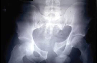
Is putting a binder around the greater trochanters and pulling going to help? Will it produce anatomical realignment? I think you will agree that it is hard to know. In this case it might rotate the left hemipelvis inward and create even more distortion. You might also guess that some traction on the left leg before you apply the binder might get a better result too. More on this later.

Is there any actual evidence that things can get worse with a binder in vertical shear? Tan’s paper had six of this injury type. Two of the six had a fall in MAP immediately after the binder was applied, one by 20mmHg! It is a bit crazy that we are discussing studies with six patients but this is the level of published evidence to date. Such as it is, the evidence is that one in three vertical shear injuries deteriorated immediately after the binder was placed. Toth’s paper found that 14/17 patients had improved alignment post binder in this group so it often does some good. Unfortunately you have to think really carefully about this group, and be prepared to loosen it off again if you don’t get the response you were hoping for.

Yes, loosen it if the patient deteriorates! Primum non nocere. Remember there is as yet no study that has shown significant mortality improvement with pelvic binders. They are not a standard of care. If what you do makes thing worse then backing off is the right thing to do. I try not to let my own psychological need to do everything I can for the critically injured patient in front of me drive me to do things that might actually harm the patient. Sometimes less is more.

So what are we left to conclude?

  • AP compression – makes biomechanical sense and low level evidence binders help
  • Lateral compression – makes no biomechanical sense and real world evidence binders increase fracture displacement. Is “just holding it still” enough?
  • Vertical shear – a really difficult group; evidence of haemodynamic and anatomical improvement in the majority but clinically significant deterioration has also been documented
  • The real world as always is a bit more complex than this and mixed injury types happen
  • And of course, no evidence yet overall that binders have a significant effect on the outcome that matters in this case –mortality.

It should be pretty obvious that the type of fracture should be the guide to whether or not a binder might help. This is great if you are doing an interhospital transport and have an Xray. Not really helpful though if you are at the roadside, on an oil rig, or at a remote clinic 1000kms from the nearest trauma centre with no imaging (as our teams frequently are). So how can you work out whether a binder will help?

First thing is reading the injury mechanism. If you are at the scene you may get a lot of clues about the force vector, particularly in motor vehicle trauma. This is a photo of an incident I attended a few months ago.

Powerpole copy

In this case the car had slid sideways into the power pole striking the driver directly in the right side with such force that she had broken the centre console with the left side of her pelvis and was partially in the passenger seat. This can only be a lateral compression injury and there is no way a binder can help. Direct frontal injuries are also pretty obvious and the injury type is going to be an AP compression if a pelvic ring fracture is present.

This is good as far as it goes. It really does not help much with other mechanisms like pedestrians and motor cyclists. Were they side-on or front-on when they were hit by the truck? Motor cyclists can have a significant rotational component to their flight before they hit something which can make prediction of injury patterns really problematic.

There is one other trick which can give you a really valuable clue. Symphyseal diastasis is the hall mark of the AP compression injury. This is the sign that the “book is open”. If you can identify this then you can identify the group that is likely to benefit from a binder to “close the book” (although some will have vertical shear so care is still required). This is yet another use for my trusty companion, the handheld ultrasound.

The width of the pubic symphysis can easily be measured with the same high frequency linear probe that you use to exclude a pneumothorax. The upper limit of normal width measured at the point shown in the image is <25mm in adults (Bauman). I am not aware of any published data on children. As with all things there is a bit of variation here and cadaver studies have shown that anterior sacroiliac ligament disruption is likely for displacement greater than 45 mm and unlikely for values less than 18mm. So if the symphysis is less than 18mm you can be very confident the pelvis is not “open”.

Ultrasound copy

Clinical ultrasound copy

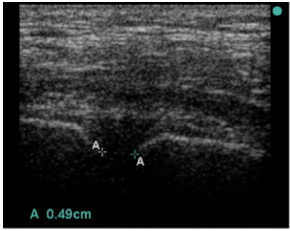
Note that in the source study for the reference range they failed to achieve a measurement in one case because the symphysis was wider than the width of their probe. You may have to move the probe from side to side to pick up both sides of a really wide symphysis.

If the patient does not have symphyseal widening on the other hand there is no reason to believe that a binder will help and they may well have an injury type that will be worsened by a binder (the symphysis does not open in lateral compression). Ultrasound is likely to be our best guide as to which patients have the possibility of benefitting from a binder whilst avoiding those where harm is the more likely outcome.   Some patients with vertical shear and an open symphysis may still deteriorate so there is no guarantee, but ultrasound will at least allow you to identify the group who have the possibility of benefit rather than harm.

As with so many things in prehospital care we need some good studies in this area. In the meantime, read the mechanism, read your ultrasound screen and be judicious in applying binders. Harm has occurred with these devices – they are not a universal panacea. Much of the art of medicine is picking the right patient for the intervention so you maximise benefit and minimise harm. This patient group is no different.

And thanks for the comments. Julian Cooper’s thoughts helped me work through my own theories on the issues and I have realised that our theories and the observational evidence don’t seem to align. There is also some potential new approaches to the massively haemorrhaging pelvis that are easily applicable in the prehospital environment and those are worth looking at too.

So looks like I am doing part 4. Stay tuned folks.

Bauman M et al. Ultrasonographic determination of pubic symphyseal widening in trauma: the FAST-PS study. The Journal of Emergency Medicine, Vol. 40, No. 5, pp. 528-–533, 2011.

Doro CJ et al. Does 2.5 cm of symphyseal widening differentiate anteroposterior compression I from anteroposterior compression II pelvic ring injuries? J Orthop Trauma. 2010 Oct;24(10):610-5. doi: 10.1097/BOT.0b013e3181cff42c.

Tan ECTH, et al. Effect of a new pelvic stabilizer (T-POD1) on reduction of pelvic volume and haemodynamic stability in unstable pelvic fractures. Injury (2010), doi:10.1016/j.injury.2010.03.013

Toth L, King KL, McGrath B, Balogh ZJ. Efficacy and safety of emergency non-invasive pelvic ring stabilisation. Injury, Int. J. Care Injured 43 (2012) 1330–1334

The Bind About Pelvic Binders (Part 2)

This is part 2 in Dr Alan Garner’s series on pelvic fractures and the approach to binders. You can find part 1 here

In part one we had a look at the evidence for benefit from pelvic binders. In short there is no study yet published showing a significant improvement in mortality. Not even a cohort study.

Of course, it still might be OK to use them if they possibly help as long as there is no evidence of harm either (and they don’t cost too much). The probability of good has to outweigh the probability of evil. It is the potential for evil that I want to examine now so we can see where the balance lies.

Before we can do that though we need to have a quick look at the types of pelvic ring fractures (no one is suggesting that non-pelvic ring fractures of the pelvis benefit from a binder). So sorry folks but we have a bit of theory to re-visit.

Forces Down There

I use the Young and Burgess classification system as it is based on the force vector that caused the injury. In the prehospital world mechanism of injury is almost the only guide to injury type that is available to us (ultrasound may also give us some clues but we will talk about that in part 3).

AP compression injuries

AP Compression copy

This is an anteroposterior (AP) compression injury. This is the kind of fracture you see in frontal motor vehicle collisions, commonly in motor bike riders, and people who have been crushed by a vehicle rolling over their pelvis for example. The hallmark is pubic diastasis with or without disruption of the SI joints. The AP compression causes the pelvis to open: one or both hemipelves undergo external rotation.

External rotation of the hemipelvis results in an increase in the volume of the pelvic cavity which then allows more pelvic haemorrhage to occur before the osseous and soft-tissue structures cause tamponade. Exsanguination is the primary risk & reduction of the increased pelvic volume is one of the goals of prehospital care.

When I was a boy Master taught me the way to reduce a fracture is to reverse the force that caused it in the first place. With this type of injury a pelvic binder makes biomechanical sense because it reverses the direction of the force which caused it. In severe AP compression injuries one or both hemipelves have been rotated backward. Applying a binder will rotate the hemipelves back towards each other, or “close the book”.

Book copy

As I mentioned in part 1 there is very little evidence on whether this is actually helpful despite the theoretical benefit. Tan’s study was observational and involved only 15 subjects in an emergency department setting. All subjects had been X-rayed prior to application of the device so the type of injury was known (unlike our context in most cases). Nine of the 15 patients in this study had AP compression type injuries with wide diastasis of the pubic symphysis.   Although there is some missing data, all patients with this pattern either had no change in MAP or it improved. So far so good.

There is a similar English study with 3 severe AP compression injury patients who improved with a binder (Nunn) but numbers are obviously pretty small.

Croce’s study appears to have had mostly AP compression fracture types (186 patients with breakdown between types not stated). Decreased transfusion requirements were found in the binder group at 24 and 48 hours (significant), the patients had decreased length of stay (significant), and lower mortality (non-significant). This does provide some support for use in severe AP compression injuries noting the methodology issues which I discussed in Part 1 with a retrospective study that included patients over a 10 year period.

There are a number of other studies which show improved alignment +/- blood pressure rise in AP compression type fractures in trauma patients, in cadavers and even in one prehospital study. None of these studies assess patient outcome though (I acknowledge this is difficult in cadaver studies!) Reduction can be so good that the fracture is difficult to see on subsequent Xray.

So in AP compression injury all the evidence points to better anatomical alignment, higher blood pressure, lower transfusion requirements, and shorter length of hospital stay when you use a binder. Mortality might be better too, but this remains to be proven. The important thing is there are no reports of adverse events in this group. When you see this fracture type on Xray or the mechanism suggests this injury – go for the binder. The risk of adverse advents is certainly outweighed by the possible benefits based on the best current evidence.

Lateral compression injuries

Lateral compression copy

Lateral compression injury results in internal rotation of the affected hemipelvis. This internal rotation decreases rather than increases the pelvic volume so they tend to bleed less than the other types. Life threatening haemorrhage is still possible though. The hallmarks include sacral buckle fractures and horizontal pubic rami fractures.

Remember my boyhood teaching – “Grasshopper, to reduce fracture you must reverse force that caused it”. There is an obvious problem here as applying a binder replicates the causal force and if anything is likely to make it worse.

Have a look at this Xray of a lateral compression injury. Put a binder around the greater trochanters and pull. Are you a force for good or evil?

X-ray copy

So what is the evidence? The Tan paper did not include any lateral compression injuries – remember that they had looked at the X-ray prior to application. I assume they looked and thought “well that is not going to help”. There is no evidence the Croce study included any either.

Is there evidence that a lateral compression fracture can get worse with a binder? (You have to be suspicious when binder studies appear to have avoided this fracture type altogether).

A recent Australian study (Toth) from 2012 had 8 cases with lateral compression that had binders applied. In three it resulted in increased pelvic deformity on subsequent Xray. They did not report the haemodynamic consequences. In the other 5 there was no improvement. There is biomechanical evidence of this in cadavers too e.g. Bottlang et al (if you look at this paper note again that they did not even attempt it in the LC3 injuries – the most severe grade).

Now this really disturbs me. There are docs I have met who are adamant that pelvic fracture patients should not be logrolled & should only be moved on scoop stretchers etc because the fracture fragments might move just with this limited motion. These same docs are however happy to put a binder on regardless of mechanism and pull, creating a much larger force than a logroll does, when we have direct evidence that binders increase fragment displacement in lateral compression injuries. Some consistency would be nice.

The bottom line is that there is no theoretical reason to believe that binders help in lateral compression injuries and lots of reasons to think they might make things worse. There is direct evidence in real world trauma patients that increased deformity of the pelvis does occur. There is no published data at all on the haemodynamic consequences when this happens, but I am betting you are not going to see improvement. The balance of risk here is on the dark side, not the light.

Bottom line is leave the binder in the bag in the bag for clear lateral compression mechanisms. It cannot help and there is published evidence of harm.


(Stay tuned for part 3 where we’ll get to vertical shear injuries – and other stuff).

References:

Croce MA, Magnotti LJ, Savage SA, Wood 2nd GW, Fabian TC. Emergent pelvic fixation in patients with exsanguinating pelvic fractures. Journal of the American College of Surgeons 2007;204:935–9. [discussion 40–2]

Tan ECTH, et al. Effect of a new pelvic stabilizer (T-POD1) on reduction of pelvic volume and haemodynamic stability in unstable pelvic fractures. Injury (2010), doi:10.1016/j.injury.2010.03.013

Nunn T, Cosker TDA, Bose D, Pallister I. Immediate application of improvised pelvic binder as first step in extended resuscitation from life-threatening hypovolaemic shock in conscious patients with unstable pelvic injuries. Injury, Int. J. Care Injured (2007) 38, 125—128.

Bottlang M, KriegJ C, Mohr M, Simpson TS, Madey SM. Emergent management of pelvic ring fractures with use of circumferential compression. J Bone Joint Surg Am 2002;84-A(Suppl 2):43–7.