Category Archives: Prehospital Trauma

Blood Warmers (Sort Of) In The Wild

This post (after, let’s face it, a massive break) is the written version of a talk by Andrew Weatherall for the Aeromedical Society of Australasia Conference 2019, held in Perth.

 This is a thing that’s about blood. And the roadside. And the lab bench. Naturally it starts with a story because clinical research should start with a patient. That’s why we do it. It’s a really topic to talk about because the management of transfusion, and the questions it brings up might seem simple at first glance. But a transfusion in the prehospital environment is a particular example of people across a whole system working for a patient.

And the questions clinicians ask that start with something small are the sorts of questions that make a difference in simple ways to every single patient.

A Mountain Story

You have to cast yourself back to the late 90s. Back when the millennium bug was a spooky tale and not an impressively well managed systems vulnerability.

On a particular morning back then there was a car accident in the Blue Mountains, just outside of Sydney. It was serious. The driver had died.

These were the times when asking for a team with a few more options waited until the ambulance officers had got to the scene and started working very hard. So around 28 minutes after the accident, the CareFlight crew on that day got a call to attend to try and help a passenger. Around 52 minutes had passed by the time they reached the patient.

In that time a lot of difficult treatment and rescue work had already happened. The patient, a 15 year old was accessible but still a bit stuck. The paramedics had already started care. In this case this included a large bore cannulae and 6 litres of polygeline fluid resuscitation.

I should have mentioned it was 7 degrees Celsius this particular morning.

Over the course of the extrication all the crews on scene combined. The patient was intubated because the combination of their injuries, diminishing conscious level and the respiratory rate of 40/min suggested something was up.

They received the 4 units of red cells that the CareFlight crew was carrying. The heart rate of 140/min and blood pressure of 70 mmHg made it clear bleeding was an issue. It was an issue that needed more red cells. How does that happen on a cold day in the mountains in 1997? The local hospital, nothing like a trauma hospital sends O negative red cells when they receive a call. The police deliver it.

The patient had a chest drain inserted after their breath sounds changed unilaterally.

But it was clear they needed more fluid resuscitation. And more.

In total, by the time the patient reached their destination hospital a flight later and about 132 minutes after their injury, the combined teams had managed to find a total of 15 units for that patient. Which gave the hospital a chance to get working.

The receiving hospital that day was Nepean hospital, at the foot of the mountains (and these days not a major trauma centre). They received a critically unwell patient and had a long day in the operating theatres. A long day with a long list of injuries who received a further 56 units of red cells, 16 units of platelets and 19 units of FFP.

And survived.

I know they survived because they provided consent for the first case report of massive transfusion in the prehospital setting that Rob Bartolacci and Alan Garner wrote up in the MJA in 1999.

Questions

That story could be seen as an extraordinary success. It didn’t stop the crew asking questions though. This was of course before we knew nearly as much about trauma and bleeding and coagulopathy. I mean we knew it was an issue but Brohi et al hadn’t published that excellent work showing a rate of coagulopathy of 24.4% or the huge import of coagulopathy when it came to mortality.

We didn’t have nearly as much understanding of the ills of acidosis, coagulopathy and hypothermia. We didn’t even have as much evidence about how easy it is to cool patients with cooled fluids as we now do.

We knew hypothermia was bad though. So when that patient arrived to hospital with a temperature of 29.5 degrees Celsius and a heart rate of 80/min, the crew started thinking about ways to do it better.

These days of course we prioritise haemorrhage control. We have a different approach to administering massive transfusions. We’d be reaching for tranexamic acid. Along the way though the first question was ‘how do we try and make our fluids warm?’ Red cells come out of the esky at around 4 degrees Celsius. Patients are not designed to meet that temperature halfway.

Lots of things have been tried over the years. In the past we’d relied on the Australian sun. We’d relied on the toasty armpit of an emergency service worker probably wearing a non-breathable fabric blend that isn’t very flattering to the profile. We’d tried gel heat pads that you’d use to ease a muscle ache.

Fast forward a decade and a bit though and we finally had a portable fluid warming device small enough to help us out. We felt pretty good about the Belmont Buddy Lite too. It was a huge step up, we figured, from what we’d been doing.

Clinicians still ask questions though. This time it was another CareFlight specialist, James Milligan, who started asking questions. It was a pretty simple one really. ‘Maybe we should check how much better it is than all the other options we’ve tried?’

Simple, right?


Setting the Right Rules

Now one of the great challenges when you’re trying to do bench testing of a device for prehospital and retrieval medicine is getting the balance right. You want to produce measurements that are rigorous and reliable enough to give you real information. The risk when you do that is that you do things so differently to the environment that counts for us that it no longer represents something that still applies at the roadside.

So the natural choice if you want to bench test a prehospital blood warming device is this …

… a bespoke cardiopulmonary bypass circuit to deliver reliable flows, measure the temperature at multiple sites, measure pressure changes across the circuit, collect the first unit of red cells you administer, spin it down and cool it then recirculate that blood to deliver the equivalent of a second unit.

With a plan to fix the flow rates to 50 mL/min (the suggested rate for the Buddy Lite), randomise the sequence of runs and repeat each type of run 3 times to generate useful data. We standardised as much as we could, including the spine board we used for the ‘on a warmed spine board in the sun’ group and had the board heating for a standardised period of time. We used a single armpit to generate body heating. (Yes, I can confirm that sometimes you have to sacrifice a few frozen off armpit hairs for science.) You get those gel pads ready to go. You get the support in an entirely different setting from your blood bank.

Then it’s time to test how MacGyver works in real life.


Phase 1: A Warmer vs MacGyver.

This study turned out to be one of those ones where the results match pretty much what you thought. You can find the full paper here but the key table to pull out is this one.

Even with this simple study there are a few interesting points to note:

  • There is actually more warming than you might expect when you just run the fluid through an intravenous fluid line.
  • The Australian sun actually did pretty well.
  • My armpit is just an embarrassment. Only for this reason.
  • Gel pads just don’t have the contact time to count.
  • The thing specifically designed for warming turns out to be better than warming.

Phew. Done and dusted.

Except we had more questions.

The thing is as clinicians we knew that delivering at 50 mL/min is probably not what we do when a patient is really critically unwell. There’s every chance it’s quicker. It certainly was in 1997. Maybe even pulsed because someone is squeezing it in.

So to really make the benchtop more like the place we work, we wanted to test different flow rates. We also thought maybe we should double check that putting red cells through a rapid temperature change across a small area of space where at least some pressure changes happen inside the device was still OK for red cells. What if we haemolyse a bunch of hose bendy little discs.

Happily just as we got there, more devices hit the market.


Phase 2: Warmer vs Warmer

We had some more limits to set though. The devices we’re after for prehospital work need to be light enough to not be too annoying. They have to be free of an external power source. Ideally they’d be idiot proof. I mean folks like me just a little overloaded at the accident scene need to use them.

So we looked for devices that could do the job at a total weight under 1 kg and that operated as standalone units. We ended up with 4 to test:

  • The Thermal Angel.
  • The M0
  • The Hypotherm X LG.
  • The Buddy Lite.

This time with flow rates of 50 mL/min, 100 mL/min, and 200 mL/min (the maximum rating of any device was 150 mL/min but we also knew that with a pump set we can get up as high as those numbers). Still randomised. This time with fresh units of red cells so we could test for haemolysis on the first run through the circuit.

The testing circuit was a little different this time, mostly because it did things better and would reliably deliver the flow rate all the way to 200 mL/min. It was a bit quicker to cool the units too.

The results this time are best shown in a couple of the tables. At 50 mL/min there’s one warmer that’s clearly performing better than the others. The red cells reach the thermistor that reflects delivery to the patient at 36.60C. The best of the others only gets to 30.50C.

When you get to higher flow rates the difference is even more marked.

That same device gets the blood to 32.50C. The others? 23.7, 23.5 and 19.40C.

That is a heck of a difference.

Along the way we couldn’t pick up any evidence of haemolysis. We count that as the best sort of negative result.

Oh, that warmer was the M warmer. We switched.

The Deeper Bits

A study that started with a very simple question ended up being a pretty fun excursion into the lab. Except for that one thing where the clamp went on the wrong bit of the circuit.

For no reason at all I’m just going to mention that wearing PPE not your own clothes is something you should be glad you do at work. It pays off.

The other thing worth noting is that all of these devices go through a process before they become available for sale commercially. That is part of what the TGA does. And they all do kind of what they say on the box. They do warm things up. They are unlikely to cause problems. They’re not the same though. You could even argue that some of them performed so below the level of the most effective device we tested that it’s only marginally better than our next reference, my armpit.

Everything that goes to market has its own story, just like the little pig who goes to market I guess. The TGA is obviously very rigorous in applying its incredibly voluminous guidelines. And I wouldn’t suggest that the manufacturers or those sponsoring the devices t get into the market haven’t likewise followed every part of that process.

What is less clear to me is how you demonstrate ‘suitable for intended use’ which is one of the essential principles that must be shown to be met. Near as we can tell there wasn’t prior testing of these units that so rigorously reflected how we’re likely to use the units in our actual practice prehospitally.

There is a requirement to show that the design meets appropriate standards and that there is clinical evidence. However you are permitted to show that testing is similar enough to the area you reckon it can be used that it gets signed off. On my read of the guidelines you can even show good evidence for the principle of blood warming and that a similar device has been shown to do that well and say ‘our tech specs show we can do the same thing so tick here please’. Extrapolating practice from the hospital or other settings to prehospital and retrieval medicine is a thing we often have to do but that doesn’t mean we should just accept that.

So when you look at a design like, say, the Buddy Lite (a device that has served us really well and that was a huge step up from what we were doing) you can see that it would meet lots of essential principles about not exposing the patient to harm, being safe to handle and operate and warming the infusate. As long as it is delivered as suggested by the manufacturer at 50 mL/min.

And that’s the bit of evaluation that we can’t rely on the TGA to do for us. Is 50 mL/min what we’re after?

We need clinicians asking questions.
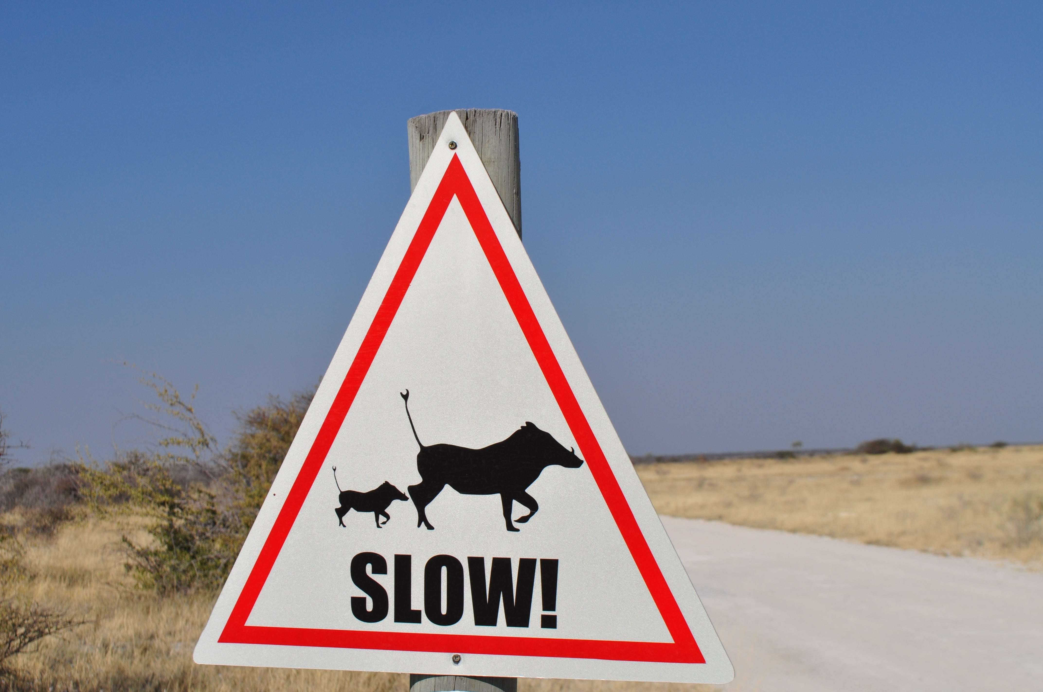
Slow Down

So we started with a question. Actually the questions have been happening since at least the 90s.

Simple questions asked by clinicians thinking about the patient in front of them are really useful. They take you in unexpected directions. They lead you to work in teams just like you do at the accident scene. It’s just that this team involves some doctors and a paediatric perfusionist and a haematologist and a lab technician and the blood bank and the Red Cross and a statistician in Hong Kong.

And we’ll keep asking questions. Along the way on this study we figured out that the Hypotherm was just a little challenging to use. In using the M warmer we’ve picked up ways we need to manage the battery recharging.

Clinical teams are vital to doing things better for our patients not just because we actually do the doing, but because the studies that get out there need to be interpreted by people who know the operating space. Or it needs to be clinical teams and patients inspiring studies in the first place.

Someone needs to keep an eye on the pigs when they’re trying to get to market.


The References Bit:

OK that’s quite long.

That case report is this one (and obviously the patient has given permission for its use in contexts like this):

Garner AA, Bartolacci RA. Massive prehospital transfusion in multiple blunt trauma. Med J Aust. 1999;170:23-5.

The first of the blood warming papers is this one:

Milligan J, Lee A, Gill M, Weatherall A, Tetlow C, Garner AA. Performance comparison of improved prehospital blood warming techniques and a commercial blood warmer. Injury. 2016;47:1824-7.

The more recent one comparing devices is this one:

Weatherall A, Gill M, Milligan J, Tetlow C, Harris C, Garner A, Lee A. Comparison of portable blood-warming devices under simulated prehospital conditions: a randomised in-vitro blood circuit study. Anaesthesia. 2019;8:1026-32.

If you want to read that Brohi et al paper again it’s here:

Brohi K, Singh J, Heron M, Coats T. Acute traumatic coagulopathy. J Trauma. 2003;54:1127-30.

You might like to reflect on just how quickly you can cool someone down with cold crystalloid:

Kämäräinen A, Virkkunen I, Tenhunen J, Ali-Hankala A, Silfvast T. Induction of therapeutic hypothermia during prehospital CPR using ice-cold intravenous fluid. Resuscitation. 2008;79:205-11.

If you have lots of spare time you might like to read the TGA regulations. (Note they are being updated.) I advise drinking coffee first.

And did you get all this way? Then you definitely need to watch this and relax.


Making Things Clot

It’s been a solid break but here is Andrew Weatherall returning with a bit about just one approach to getting access to plasma in the field. 

In prehospital medicine there aren’t really many limits to what we can carry. Ultrasounds are routine. Intubation gear is boring. We even carry patients some of the time.

Plus since 1987 we’ve carried red cells for transfusion. Which is excellent. And we’ve used plenty. Sometimes though you want to change things up and changing things takes time. Like say, changing what you carry for transfusion. Which only took us 30 years or so.


How you get it done…

Us carrying red cells has been reality through a range of different attitudes to transfusion. Where we find ourselves now of course is thinking that if you need fluids in the setting of trauma then the fluid should not be see through. For a while though we’ve been trying to figure out how to extend beyond red cells.

The thing is that now that we know that acute traumatic coagulopathy (ATC) is a thing, and we’ve known that for a pretty decent amount of time now, you need to be wrestling with how to try and deal with the coagulopathy bit, not just dish out the vampire-sating stuff.


So what to use…

If you go to look at the literature, for a while there’s been plenty out there saying that fibrinogen might be a useful thing to go with. So we’ve been waiting a while for freeze-dried cryoprecipitate but last time we looked it came out as a little bit pricey for our taste but there was something more significant.

Time.

djim-loic-69263-unsplash
Blurry, blurry time.

The more significant thing for our work profile was just the fact that preparing FCCs takes a bit of time and would probably take a team member out of action for too long. That might be a different story if we were doing retrieval or more remote pick-ups, but for metropolitan responses it just didn’t quite seem to fit.

So we kept giving the red cells. Then when TXA gained some evidence, we added that. That one’s quick.

Trauma centres kept publishing on the role of viscoelastic testing but that didn’t fit our work profile either. So we were stuck there.

And whole blood was not on the table in our neck of the woods.

It kept nagging at us though. What’s the other thing we should be giving?

Which is why we went to chat to a clever person at the place we pick up our blood.


The Ideas Bank

At this point something we’d discounted as an option suddenly became an option. Plasma.

It just hadn’t seemed like an option for us because of the issues around thawing time and the need to use it within 4 hours. That’s not true anymore though. Extended life plasma (ELP) is now available and is OK to use for 4 days after thawing. Add that to some evidence that group A ELP is suitable for use as a donor unit in the adult patient population (rather than having to always use group AB) and our blood bank contact had come up with a plan to do something different.

So this was the plan:

  • Switch what was in our blood esky from 3 units of O- red cells to 2 units of O- red cells and 2 units of group A ELP.
  • At the end of the shift (this operation is running one shift per day), return the products as usual to the blood bank.
  • The ELP can then go back into the pool used by the hospital blood bank.

And we figured we’d start the plasma early, maybe after the first lot of red cells.

There were to be some limits of course, particularly when it comes to kids. Try as we might, we just couldn’t find guidance for kids under 11 to see if group A ELP alone would be appropriate. Same story for the paeds haematologists. So kids were out.


The Introduction

The whole thing got moving within a few short months and the plasma hit the esky in April. It was introduced with lots of discussion and education and an undertaking to keep track of the results.

 It really was seamless. We just sort of needed the evidence to back up the expert advice of the haematologists.


Catching Up

And this is where another bit of fortuitous timing worked in our favour. A trial came out.

PAMPer reared up in the NEJM showing a very solid difference in mortality in those given plasma in the prehospital phase. This was patients between the age of 18 and 90 and with evidence of haemodynamic things that matter – systolic blood pressure under 90 mmHg (or a single reading hitting 70 or below) and heart rate above 108/minute. 5 minutes of being arrested was also enough to get a number of people kicked to the side.

It’s also worth mentioning that they just gave their 2 units of plasma in their entirety. Some of the centres were carrying red cells so if they were being treated as recruitment centres administering “standard” resuscitation for a particular day, there was a total of 13 of the 27 centres who might go ahead and give some red cells.

A total of 501 participants were eventually randomised. 30 days after the patients were randomised the number of deaths in the standard care group was 89. This is significant compared to the 53 deaths in the plasma group. After regression analysis, those in the plasma group had a risk of death 39% lower than standard care. There were also mortality differences at 24 hours and in the “in-hospital” cohort.

As a bonus, patients in the plasma group received fewer units of blood components overall. Plus they had no documented cases of transfusion-related lung injury during the trial.

So that was enough to make us tweak things a little more and consider the plasma as our first line resuscitation in most instances, followed by the red cells.


What makes it work?

So for now this solution works in our context. That’s not to say it would work for everyone or would be the best choice for different work profiles. We note that London’s excellent HEMS service has just recently introduced bags with both red cells and FFP in them and that’s a different approach again which will be worth watching. And even more recently the crew from Kent Surrey Sussex Air Ambulance Trust have published a retrospective study sharing their success with freeze-dried plasma.

It’s also not to say we won’t seek to tweak things in the future. There are a few key things that made the use of ELP work for us:

  1. Local experts

There’s no doubt that the ‘who you know’ factor helped in setting this up. Being able to approach the local expert, in our case Dr Leo Pasalic (full credentials below), who could think laterally when we approached with a query opened up an avenue we had dismissed. Leo was also able to place it in the context of a broader understanding of the literature. There’s no literature search that’s a replacement for long experience and cultivated expertise.

  1. A very excellent blood bank

A nearby blood bank that supports that expert person with great service is a pretty crucial part of the mix. As clever as Leo is (and he is pretty clever), the whole plan hinges on other clever people who pull off the ‘let’s make this happen’ bit and have that extra understanding of the science and logistics. For us those other key people were Dr Kifah Shahin and Hayley Keenan (you should check their full credentials below too). From the time of deciding to do this to actually carrying the ELP was weeks at most and the logistics have never been an issue. They added the packs and made it work.

  1. Reduce, reuse, recycle

When giving blood products we are, naturally, also trying to nourish our inner greenie. I mean, as it is we’re aiming to reduce the overall usage of blood products throughout the admission. We also want to make sure we’re not thawing the ELP and not being able to use it. Blood products are a precious resource and the cost of production is also high. To date, not a single unit of ELP has gone unused. It’s thawed for the prehospital service, returned at the end of shift and invariably used well within the permitted timeframe by the very big hospital for other patients.

gary-chan-351213-unsplash
This is a terrible place for blood products to end up.
  1. The follow-up plan

The plan for tracking the experience of use is already in place so we can confirm our local experience with this new option. Trial evidence is great but ongoing real world monitoring has to come with the territory.

We’ve come a fair distance from our early forays into prehospital blood administration which involved the side of a road, a hospital somewhere nearby and police doing high speed runs to deliver those units.

It’d be good if the next step took less than 30 years though.


Notes:

Before we get into the references, we would really like to acknowledge the support of the NSW Health Pathology through their Blood Bank at Westmead Hospital and the brilliant staff working there.

Dr Leo Pasalic is a staff specialist in laboratory and clinical haematology and transfusion medicine and gets to cover the medical supervising responsibilities at the Blood Bank at Westmead Hospital.

Dr Kifah Shahin is the Transfusion Lead Scientist for the West, Rural and Regional laboratories in NSW Health Pathology.

Hayley Keenan is a Transfusion Senior Scientist at NSW Health Pathology Westmead.

That Blood Bank they work at is part of the Department of Haematology at the Institute of Clinical Pathology and Medical Research, NSW Health Pathology, Westmead Hospital. The hospital has been on that location for 40 years. The ICPMR got there first, having been on the site for 41 years.

Now, the reading list.

Here’s the link to the PAMPer thing again:

Sperry JL, Fuyette FX, Brown JB, et al. Prehospital Plasma during Air Medical Transport in Trauma Patients at Risk for Hemorrhagic Shock. New Engl J Med. 2018;279:315-26.

This isn’t the only story out there of course. The paper from Moore et al is worth a look.

Moore HB, Moore EE, Chapman MP, et al. Plasma-first resuscitation to treat hemorrhagic shock during emergency ground transportation in an urban area: a randomised trial. Lancet 2018;392:283-91. 

And that Kent paper again:

Oakeshott JE, Griggs JE, Wareham GM, Lyon RM. Feasibility of prehospital freeze-dried plasma administration in a UK Helicopter Emergency Medical Service. Eur J Emerg Med. doi:10.1097/MEJ.0000000000000585

Now, some others:

Winearls J. Fibrinogen in Traumatic Haemorrhage: A Narrative Review. Injury. 2017; 48:230-42.

Lance MD, Ninivaggi M, Schols SEM, et al. Perioperative dilution coagulopathy treated with fresh frozen plasma and fibrinogen concentrate: a prospective randomised intervention trial. Vox Sanguinis. 2012;103:25-34. 

Schöchl H, Nienaber U, Maegele M, et al. Transfusion in trauma: thromboelasfometry-guided coagulation factor concentrate-based therapy versus standard fresh frozen plasma-based therapy. Critical Care. 2011;15:R83. 

Shackleford SA, del Junco DJ, Powell-Dunford N, et al. Association of Prehospital Blood Product Transfusion During Medical Evacuation of Combat Casualties in Afghanistan With Acute and 30-Day Survival. JAMA. 2017;318:1581-11. 


CPR in TCA: A TLA PSA

‘Don’t compress the chest in traumatic arrest…’ That’s the narrative. But Alan Garner has questions.

Do you do chest compressions in traumatic cardiac arrest (TCA)?

Don’t be dopey, right? Compressions are not important compared with seeking and correcting reversible causes. Indeed you can just omit the compressions altogether and transport the patient without them as they are detrimental in hypovolaemia and obstructive causes of arrest, right?

I would like to work through the logic of this.  I think the nidus of an idea got dropped into a super saturated FOAMEd solution and Milton the Monster* precipitated out.  The end result might be an approach that got extrapolated way beyond the biologically plausible.

The Starting Point

First let’s try to step slowly through the logic…

  1. In hypovolaemia or obstructive causes of shock that are likely in the trauma patient (tension and tamponade) and where the patient is in PEA (preferably with good cardiac motion on ultrasound) then they might still have output but below the limits of detection except by doppler – certainly not by feeling pulses.
  2. In these patients RV filling is critically impaired and chest compressions during diastole can further impair filling.
  3. This converts the low output state into a no output state, and this has to be bad.

(There is an underlying assumption I think we’d have to agree with here – that a no output state is worse than a low output state. Pretty fair call.)

denny-luan-125448-unsplash.jpg
Here’s the bit with the leap…

This all seems to bring you to ‘don’t bother with chest compressions at all as they are detrimental in all TCA patients’. Or, ‘at least don’t do them till you have done everything else and only if you have some spare hands available’.

But I’ve certainly heard those at the extreme end of the spectrum who don’t do compressions at any point and transport the patient without them even if plenty of hands and a LUCAS or two are available.

Circling Back

Let’s get back to each step along the way.  The first point was that the patient in PEA (who has an organised rhythm and probably decent contractility on ultrasound) is producing subclinically detectable flow which we do not want to mess with.  This is great if the patient is that exact patient with PEA and a sniff of reasonable contractility (this and pupillary reflexes are the two things you really want to see in an arrested trauma patient).  No argument from me here.  But what if the patient starts in what can only be a no output state – like asystole or VF?

Should we not be trying to convert these clearly non-perfusing, no output rhythms into at least a low output state by performing CPR?  My assumption here is that it should work like conventional CPR – stop for as long as you need to perform critical interventions, but absolutely minimise hands off time otherwise.

We have already agreed that a low output state is better than a no output state and indeed this is the nidus of the argument for withholding compressions in TCA.  But we are also being asked to assume that a low output state is the most likely situation the patient has ended up in. This is the justification for withholding all compressions, on the basis that PEA might be present, and that there is a theoretical chance that RV filling might be impaired if a compression coincides with diastole.

Again let me emphasise that I am not saying that compressions should be performed at the expense of treating reversible causes like hypoxia or tension pneumothorax. These things absolutely take priority – just with minimal interruption to compressions.

What I am seeing however is intubated patients with bilateral thoracostomies and no tamponade on ultrasound who are in asystole not having CPR performed.  No cerebral or cardiac perfusion is even biologically plausible without compressions in these patients who do not have a perfusing rhythm.

To the Library

So for the assumptions that let you say ‘compressions are a waste of time’, you’d need to be pretty sure that PEA is overwhelmingly the most likely thing you’d see.

But it’s not.

The most common rhythm in TCA appears to be asystole and it amounts to over two thirds of patients. These patients are not in a low output state with critical RV filling issues. They are in a no output state. There will be no output unless you do something to encourage it.

Another 7% are in VF. This would be another rhythm where chest compressions are indicated. So that means in total about 75% of TCA patients will have a rhythm where compressions may help. So does omitting compressions for all, to address a ‘sometimes’ thing in the 1 in 4 range, seem like a sensible balancing of probabilities?

It seems more sensible to keep compressions as the default in TCA given that most patients are in a “definitely no output” rhythm. If you find PEA then it’s your call I guess. Personally I will still be doing compressions particularly once hypoxia, tension pneumothorax and tamponade have been excluded. Using an approach like the HOTTT drill this can generally be achieved within a couple of minutes of patient contact and that should hopefully keep ‘no flow time’ to an absolute minimum.

What about studies were compressions are delivered?

Well this recent study noted rising EtCO2 levels with chest compressions in TCA, which suggests some increase in flow. A recent Japanese study of 893 blunt trauma arrests who received closed chest CPR found a 28 day survival rate of 6%. In the context of TCA in blunt trauma that’s a pretty decent number.

There’s also a study from Germany where compressions are the routine which combines data from the national cardiac arrest registry and trauma registries. They looked at ROSC rates in traumatic arrest patients and the outcomes. They found high rates of ROSC with more than a quarter having spontaneous output at hospital admission, though only 7% survived to hospital discharge. CPR was the default and survival rates comparable to the best reported in the literature. It’s not nothing.

So it seems to me we can make the cognitive load of TCA patient management about the same as other forms of arrest. It can sort of just be normal ALS with a big focus on the processes to reverse the reversible, supported by something like the HOTTT drill.

Am I missing something?


Notes:

* It was an editorial decision to leave the reference to Milton the Monster there because sometimes you just have to let people show their age.

The image of that leaping reptile came from excellent sharing site unsplash.com and was posted by Denny Luan.

Now, the papers:

Leis CC, Hernández CC, Blanco MJ, et al. Traumatic cardiac arrest: Should advanced life support be initiated? J Trauma Acute Care Surg. 2013;74(2):634-638

Here’s the study with the CO2 stuff:

Open chest cardiac massage offers no benefit over closed chest compressions in patients with traumatic cardiac arrest.  Journal of Trauma and Acute Care Surgery: November 2016 – Volume 81 – Issue 5 – p 849–854 doi: 10.1097/TA.0000000000001227 

Here’s the Japanese study mentioned:

Comparative Effectiveness of Emergency Resuscitative Thoracotomy versus Closed Chest Compressions among Patients with Critical Blunt Trauma: A Nationwide Cohort Study in Japan.  Suzuki K et al. PLoS One. 2016 Jan 14;11(1):e0145963. doi: 10.1371/journal.pone.0145963. eCollection

And the German study:

Gräsner J-T, Wnent J, Seewald S, et al. Cardiopulmonary resuscitation traumatic cardiac arrest – there are survivors. An analysis of two national emergency registries. Crit Care. 2011;15:R276. 

Where’s Wally? Finding that Patient

Having advanced teams is no damn good if you’re not doing your best to get them to the patient where they might add value. Dr Alan Garner returns with reflections on recent publications on this coming out of Europe. It’s a bit of a passion of his. 


Advanced capability prehospital medical teams may well be great, but they are an expensive resource that needs to be matched to the patients that are most likely to benefit. But these patients represent a very small percentage of the total numbers of calls to ambulance services so a way of accurately filtering the calls is critical to optimising the utilisation of such teams.

This of course means an accurate case identification system is required to dispatch these teams to the right patients, and preferably only the right patients. This is a kind of ‘Where’s (severely injured) Wally?’, all day, every day.  It is really hard to pick which of the red striped shirts is blood and the signal to noise ratio about the same as a Where’s Wally picture.  In NSW only about one in every 250 calls to Ambulance is a severely injured patient.

When it comes to dispatch of advanced capability medical teams (rather than which patient should go direct to a trauma centre – the two things are not necessarily the same) there is not a huge amount of literature out there yet.  This has been identified as a priority research area for HEMS.

Wally
Even with the empty seats, it’s hard work.

Hence I was really interested in a new paper just published by the people from EMRS Scotland on case identification in severe trauma.  Those who follow The Collective will be aware of my interest in this area from the work we have done in dispatch in NSW that arose as a spin off from the Head Injury Retrieval Trial, particularly in children.   You can find an earlier post on this here.

What the Scots did was move one of their clinicians who work on the service, either a paramedic or retrieval practitioner, into the control centre to look for cases that might benefit from an advance medical team response.  Prior to this move the case identification was done by non-clinical dispatchers with some oversight from paramedics and nurses who were not involved in provision of the Scottish retrieval service.   They used a simple before and after methodology to see how many of the severe trauma cases that occurred in Scotland were identified by the new system including the EMRS clinician versus the old system with the control room staff only.

The result was an improvement in sensitivity for major trauma that increased from 11.3% to 25.9%. Although 25.9% does not sound great it is possible that the new system identified almost all the severe trauma that was in the areas that the EMRS responds to.  EMRS are not dispatched to urban areas in close proximity to major hospitals but injuries occurring in these areas were not excluded from the analysis. Since Scotland has a predominantly urban population it is very likely that most trauma occurs in urban areas (like it does in NSW) so if they are identifying a quarter of all the severe trauma cases this may represent almost all the severe trauma that is in their response area.  Unfortunately this is not examined in the paper (I mean you can’t always cover everything) and more studies will be needed to clarify this.

Regardless of this methodological issue they more than doubled their case identification rate by putting a member of the EMRS team in control of identifying the cases.  Such systems are common in the UK.  As far as I am aware the first to publish this were London HEMS in early 90s where they were able to demonstrate a really dramatic improvement in sensitivity when they put one of the flight paramedics into the central control room. So this is not a new bit of learning. It’s reinforcing what we should know.

Stories we have heard before…

When we started the Head Injury Retrieval trial 13 years ago we had something like the London system in mind.  By accident we stumbled upon an improvement though.  Between 1989 when London HEMS set up their system and 2004 when we were planning the trial the internet had arrived.  We were able to build a system where the crew at the helicopter base was able to screen the calls and identify the cases directly from the Ambulance computer system rather than putting a flight paramedic into the control centre.

This contributed to the trial HEMS system being the fastest reported to date in the world medical literature.  We could get airborne about 3 minutes faster than the reports out of London and we’re pretty confident this was related to the ability to do multiple things simultaneously because it was all happening on base. At the same time as a clinically involved crew member was looking at the details of the case, aviators could start identifying potential landing sites and making plans. The pilot could head to the helo and start the checks even as that was happening. An experienced crew of 4 looking at cases also allows plenty of bouncing things off each other. There’s just a bunch of seemingly little stuff you can start working on that adds up to a significant bit of time-saving.

The trial system was however shut down at the end of the trial in 2011, and dispatch in NSW is now done by control room staff who are not involved in service provision.

It is worth noting that the system used to identify severely injured children when HIRT was recruiting was associated with zero safety incidents of even a minor nature, no unintended dual responses by physician teams and zero cost.  But it doubled the rate of identification of severely injured children for physician team response and decreased time to a paediatric trauma centre by half an hour.

It is now seven years since this case identification system was discontinued by the powers that be and there is still no suggestion that it will be recommenced. This is despite the mounting evidence of system deterioration and concerns about inevitable missed cases and delayed responses resulting in poor clinical outcomes.

Meanwhile, elsewhere…

It is notable that other services are now commencing the direct screening by HEMS crew case identification system.  Have a look at this article on the Great North Air Ambulance in the north of England. It sounds like exactly the process we used during the Head Injury Trial to identify severely injured children in Sydney more than a decade ago.

This comment from Andy Mawson, Operations Manager for Great North Air Ambulance is central to the whole thing:

“It’s an extra set of eyes to make sure we are getting to the right patients in the fastest possible time. Essentially we’re working in support of the teams within the NWAS control centre, it’s a great example of collaborative working.”

The system used to identify severely injured children during the head injury trial in Sydney was collaborative too, not competitive.  There were extra eyes looking for the same cases rather than one set of eyes looking at the whole state of NSW trying to find severely injured Wally across four different control centres.   How can this not be better?

But this is a refinement of the system the Scots studied; putting case identification into the hands of clinicians that provide the services is the core of the system. Why does this work?  I think the clinicians directly involved in provision of advanced prehospital teams just understand the services they are dispatching better.  They understand the actual logistics of response and the capability that can be delivered on scene.  It seems that you need more than a set of guidelines to do this sorting quickly and effectively.  The “gestalt” that comes from actual provision of these services is required to lift the dispatch system to the next level.

Sydney previously had a case identification system that was world leading and is now being replicated in other places like the north of England.  Sydney also had the fastest physician staffed HEMS yet reported in the world literature to go with it.

But the case identification system was switched off and the effective service area of the HEMS halved.  Missed cases and delayed activations occur frequently.

So after all this, the same questions from my last post on this topic in August 2016 still apply:

“You might ask why such a hugely effective system was discontinued in the first place.

You might ask why it has not been reinstated given the subsequent evidence.

And they would be very good questions.”


[Sound of silence].


Notes:

We are always interested in people’s clever thoughts on tricky topics. So hit those comments if you have things to say.

Now, the first of those papers on the paediatric tasking happening with the crew watching the screens is this one:

Garner A, Lee A, Weatherall A. Physician staffed helicopter emergency medical service dispatch via centralised control or by crew – case identification rates and effect on the Sydney paediatric trauma system. Scand J Trauma Resusc Emerg Med. 2012;20:82. 

The follow-up which looked at the before and after state of play was this one:

Garner AA, Lee A, Weatherall A, Langcake M, Balogh ZJ. Physician staffed helicopter emergency medical service case identification – a before and after study in children. Scand J Trauma Resusc Emerg Med. 2016;24:92. 

That paper on priority areas for HEMS research is this one:

Fevang E, Lockey D, Thompson J et al. The top five research priorities in physician pre-hosopital critical care: a consensus report from a European research collaboration. Scand J Trauma Resusc Emerg Med. 2011;19:57.

The Scottish paper (which is not at all like the Scottish play) is this one:

Sinclair N, Swinton PA, Donald M, et al. Clinician tasking in ambulance control improves the identification of major trauma patients and pre-hospital critical care team tasking. Injury. 2018. doi: https://doi.org/10.1016/j.injury.2018.03.034

That original London was this one:

Coats TJ, Newton A. Call selection for the Helicopter Emergency Medical Service: implications for ambulance control. J R Soc Med. 1994;87:208-10. 

And that letter was by me and here is the reference:

Garner A. Pre-hospital and retrieval medicine clinical governance in Sydney and the inconvenient truth. Emerg. Med. Australasia. 2017;29:604-5. 


Just because you can …

With a couple of new papers landing that touch on the issue of how you provide and measure quality care around airway management, Dr Alan Garner returns to point at big animals that are bad at hiding.

Two new airway papers have come across my desk in the last couple of weeks and I now wish I had waited a bit longer before putting up the last post on first look intubation as a quality measure.

So where to start? Well how about a place where everything is apparently big? Yes, there’s a bit of work just out of Texas which sheds further light on that first look intubation story so that’s where we’ll land.

Chasing Quality

It sounds like they have used RSI for a while but undertook a quality improvement project to try and reduce their peri-intubation hypoxia rate.  The project involved introducing a bundle of interventions described in the paper as “patient positioning, apn[o]eic oxygenation, delayed sequence intubation, and goal-directed preoxygenation”.

The paper provides copies of the protocol for intubation pre- and post-bundle intervention in the on-line appendices so I might just go through them here to see what they did differently.

The first thing is there was an emphasis on positioning in the bundle, specifically head up a bit and ear-sternum positioning.  Lots of goodness here that I strongly support.

The second measure they mention was apnoeic oxygenation.  However looking at the pre- and post-bundle policies it is evident that they used it in both time periods.  In the before period it ran at 6L/min till the sedation was given then it was turned up to 15L/min.  In the post period however it was run at “MAX regulator flow” after the ketamine was administered.  I don’t know about the O2 regulators in Texas but to me this does not sound like they changed anything significant.  I will come back to apnoeic oxygenation later.

For pre-oxygenation in the pre- bundle period they used a NRB mask (with nasal prong O2 as above) in spontaneously ventilating patients (and arrested patients were excluded) but in the post- period the pre-oxygenation had to be by BVM with two handed technique to ensure a tight seal plus PEEP.  More goodness here that warms my heart.

Delayed sequence intubation in this study refers to administering 2mg/kg of ketamine then maximising preoxygenation for at least 3mins prior to administration of the muscle relaxant.  I don’t think this is necessary in all patients but this was the policy in the bundle.

The last thing they did was “goal-directed preoxygenation”.  This refers to having a SpO2 target >93% for at least 3 minutes during the pre-oxygenation phase after the ketamine had been administered.  If they could not achieve >93% the patient was managed with an LMA or BVM and transported.  I think this represents sensible patient selection in that it removes the high risk of desaturation patients from the process.  When you look at the results you need to keep this patient selection in mind. However I agree that in their system this is a reasonable approach to ensure patient safety for which the managers should be applauded.

Show Me The Money

Yes let’s get to that money shot:

Table

I have been banging on about peri-intubation hypoxia being far more important than first look intubation rate for a while now and this data shows really clearly why.

There is no significant difference in this study in either first look or overall success rates pre and post the bundle but the hypoxia rate fell by a massive absolute 41%!  The 16% decrease in bradycardia emphasises just how much difference they made.  The managers of this system and their staff alike both need to be congratulated for this achievement as this is something that really matters.  And the first pass and overall success rates give no clue!

It really is time to drop first look as a quality measure and move on.  You could look at this paper and start wondering if it might even be worth dropping overall success rate too, which is an interesting thought.  Their policy favoured patient safety over procedural success rates by abandoning the attempt if the pre-oxygenation saturations could not be raised above 93%. It looks like it is working out well for the patients.

Oh, Back to Oxygenation

I promised I would come back to the apnoeic oxygenation issue.  I know the authors state that it was part of their bundle, but it was used in the pre- bundle period as well.  Hence there is no data here to support it’s use.

All three randomised controlled trials of apoeic oxygenation in the ED and ICU contexts (see the notes at the end) have now failed to find even a suggestion that it helps (check those notes at the end for links) and there are no prehospital RCTs.  My take is that it is time to move on from this one too and simply emphasise good pre-oxygenation and good process when the sats start to fall – or never rise in the first place like this group did so well.

Overall a big well done to the Williamson County EMS folks and thanks for sharing your journey with us.

Moving Right Along

The other paper comes out of London, where the ever-industrious HEMS group have published a retrospective review of their database over a 5 year period (from 2009-2014). They were looking for adult trauma patients they reached with an initial noninvasive systolic blood pressure of 90 mmHg or less (or where a definite reading wasn’t there, those with a central pulse only) and with a GCS of 13-15.

This gave them a total of 265 patients (out of a potential 9480 they attended). 118 of those underwent induction of anaesthesia out there beyond the hospital doors (though with exclusions in analysis they end up with 101 to look at) and the other 147 (that number dropped to 135 on the analysis) got to hospital without that happening.

Now the stated indications for anaesthesia listed are actual or impending airway compromise, ventilatory failure, unconsciousness, humanitarian need, patients unmanageable or severely agitated after head injury, and anticipated clinical course.

Now given that the inclusion criteria includes patients having a GCS of 13-15, it seems like both unconsciousness and those really impossible to handle after head injury are likely to be pretty small numbers in that 101. Even airway compromise, ventilatory failure and humanitarian need seem like they’d be not the commonest indications in that list that would apply to this patient group, though they’d account for some.

I guess it’s possible the patients were all initially GCS 13-15 on the team’s arrival but deteriorated en route, though I just can’t sift that out from the paper. Plus if that was the case it seems like you’d say that.

The Outcomes

In their 236 study patients, 21 died and 15 of those were in the ‘received an anaesthetic’ group. The unadjusted odds ratio for death was 3.73 (1.3-12.21; P = 0.01). When adjusted for age, injury mechanics, heart rate and hypovolaemia the odds ratio remained at 3.07 (1.03-9.14; p = 0.04).

Yikes, sort of.

What To Make of That? 

I guess we should make of it that … things you’d expect to happen, happen? Intubating hypotensive patients and then adding positive pressure ventilation in the prehospital setting is potentially risky for patients for a variety of known pharmacological and physiological reasons that the authors actually go into.

So the question is why embark on such a procedure where you know the dangers in detail? You’ve have to really believe in it to end up wiht 101 cases to follow up.

It feels like there’s an elephant in the room to try and address by name. I wonder if it has something to do with a practice I observed while working in the south-east of England 8 years ago. It relates to that last category “anticipated clinical course”.

james-hammond-347179-unsplash
Hovering elephant heads. They’re real.

The concept here is that if you figure the patient is going to be intubated later on in the hospital, you might as well get on and do it. Except the data here suggests that, much like you’d expect, you probably shouldn’t get on and channel your inner Nike marketing script.

Just because you can does not mean you should.  This paper really drives this home though it doesn’t really seem to come straight out and say it. It does pass the comment that “Emergency anaesthesia performed in-hospital for patients with cardiovascular compromise is often delayed until the patient is in theatre and the surgeon is ready to proceed.” Perhaps the problem isn’t using the phrase “anticipated clinical course”. It might be that you just have to remember that the anticipated course might best contain ‘risky things should probably happen in the safest spot’ in the script.

Compare and Contrast

The process of undertaking emergency anaesthesia because later the patient might require emergency anaesthesia is pretty much the complete opposite of the approach from the Williamson County EMS folks. They erred on the side of patient safety and withheld intubation if it was associated with unacceptable risk.

This paper demonstrates that emergency anaesthesia in patients with a high GCS but haemodynamic instability is associated with higher mortality.  We should probably be glad the authors have made this so apparent, because this is probably as good as we’re going to get. We’re not going to get a randomised controlled trial to compare groups. No one is allowing that randomisation any time soon making this another example of needing to accept non-RCT research as the best we’ll get to inform our thinking.

Patients with hypovolaemia due to bleeding need haemorrhage control. The highest priority in patients with that sort of hypovolaemia would seem to be getting them to the point of haemorrhage control quicker. And delaying access to haemorrhage control (because the prehospital anaesthesia bit does add time in the prehospital setting) when the patient has a GCS of 13-15 doesn’t seem to prioritise patient safety enough. Patients probably need us to adjust our thinking on this one.

That seems like common sense. The retrospective look back tells us pretty conclusively it’s a worse option for patients. And now it’s up to us to look forwards to how we’ll view those indications for our next patients. And “anticipated clinical course” probably just doesn’t cut it.


Notes:

That hovering elephant head was posted by James Hammond in a Creative Commons-like fashion on unsplash.com and is unchanged here.

How about all those things that got a mention above that you should really go and read for yourself?

Here’s that whole bundle of care paper out of Texas:

Jarvis JL, Gonzales J, Johns D, Sager L. Implementation of a Clinical Bundle to Reduce Out-of-Hospital Peri-intubation Hypoxia. Ann Emerg Med. 2018;doi:10.116/j.annemergmed.2018.01.044 [Epub ahead of print]

Those RCTs of apnoeic oxygenation in critical care environments mentioned are these ones:

Caputo N, Azan B, Domingues R, et al. Emergency Department use of Apnoeic Oxygenation Versus Usual Care During Rapid Sequence Intubation: A Randomized Controlled Trial (The ENDAO Trial). Acad Emerg Med. 2017;24:1387-1394.

Semler MW, Janz DR, Lentz RJ, et al. Randomized Trial of Apnoeic Oxygenation during Endotracheal Intubation of the Critically Ill. Am J Respir Crit Care Care Med. 2016;193:273-80.  

Vourc’h M, Asfar P, Volteau C, et al. High-flow nasal cannula oxygen during endotracheal intubation in hypoxemic patients: a randomised clinical trial. Intensive Care Med. 2015;41:1538-48.

And that paper on the hypotensive, awake prehospital patients scoring an anaesthetic is this one:

Crewdson K, Rehn M, Brohi K, Lockey DJ. Pre-hospital emergency anaesthesia in awake hypotensive trauma patients: beneficial or detrimental? Acta Anaesthesiol. Scand. 2018;62:504-14.


The Deal with Seals

Greg Brown returns to look at an important thing relevant to first responders (and lots of other people really) – the sucking chest wound. 

We’ve all been there – sitting through some kind of “first aid” training and having some kind of “first aid trainer” speaking authoritatively on some kind of “first aid style” topic. If you are like me you’ve used your time productively over the years and perfected what my wife refers to as “screen-saver mode” – it’s that look on your face that tells the instructor that you are listening intently, often supplemented by the insertion of “knowing nods” or head-tilts, but in actual fact you are asking yourself “if I was able to collect all of my belly button lint over a 12 month period and spin it into yarn, I wonder if I could make enough to abseil off London Bridge?”

Don’t get me wrong – I reckon effective and accurate first aid training should be a mandatory part of having a car / bike / truck / bus licence. More appropriately trained people should mean faster recovery rates for most injured people (and less work for overstretched first responders).

It’s just that sometimes first aid trainers teach stuff based on ‘we reckon’ or ‘that’s how we’ve always done it’ rather than evidence or knowing it works in the real world. This post is about one of those things.

“What is a sucking chest wound?”

In the Army questions come in a few different shapes and sizes. A popular one is “there is only one obscure answer you should have guessed I wanted”. Trust me, the muzzle velocity of your primary weapon is 970 metres per second.

Another popular one is “the question that should be about one thing, but is actually to demonstrate a quite tangential point”.  Like,

“What is a sucking chest wound?”

For an army instructor the answer is not what you are thinking right now. It is “Nature’s way of telling you that your field craft sucks and everyone can see you and now you got shot”.

Let’s Go With the Medical One

We’re going to go with the alternative, more medical one. A sucking chest wound is defined as air entering the thorax via a communicating wound that entrains air into the space between the lungs and ribs more readily than the lungs can expand via inspiration through the trachea.

This is about pressure differentials – in order to inhale, the lungs must generate a relative negative pressure such that air can be sucked into them via the trachea. But if you make a big communicating hole in the trachea, that might become a pretty big highway for air to enter the space with the negative pressure.

The communicating hole does need to be pretty big. Depending upon which textbook you read, this hole needs to be a minimum of a half to three quarters the diameter of the trachea. Also, the patient needs to be undergoing relative negative pressure ventilation (or, in simple terms, breathing spontaneously). If they are being artificially ventilated (which requires positive pressure) then the pressure inside the lungs will be higher than the pressure on the outside of the body; the result is that air will be forced out of the intra-pleural space (or thorax) by the expanding lung (as opposed to being entrained into the thorax via the hole in the chest).

Are sucking chest wounds really that bad?

Well, yes. They suck in fact.

A sucking chest wound creates what is known as an open pneumothorax. Let’s consider the option where that hole does not seal on expiration. We’ll get onto the also very annoying sealing with a flap version in a bit.

In this slightly not so annoying case, the patient will have a ‘tidalling’ of air in and out of this communicating hole. The effect? Respiratory compromise, increased cardiovascular effort and reduced oxygen saturations. Patient satisfaction? No, not really. Death? Maybe – depends on what other injuries exist and the ability of the individual to compensate. See Arnaud et al (2016) for more details.

But if this communicating hole were to seal itself on expiration then you now have an open tension pneumothorax. Sounds bad; IS bad.

In such a case, each time the patient breathes in they will entrain air through the communicating hole in the chest wall (that whole “negative pressure” thing in action). But when they breathe out, instead of having that additional intra-pleural air tidal outwards, the flap will seal it in place; each time they breathe in, the volume of trapped air will increase and you’ll end up with the tension bit.

How much air is required? Well a randomised, prospective, unblinded laboratory animal (porcine) trial conducted by Kotora et al (2013) found that as little as 17.5mL/kg of air injected into the intra pleural space resulted in a life-threatening tension effect.

Actually, that’s a fair bit of air…for those of you who are lazy and don’t want to do the math, that’s 1400mL for an 80kg person. But remember, any tension pneumothorax (open or closed) is progressive – each time you breathe, more air is trapped; therefore, it doesn’t take long to reach crisis levels.

“But are they common enough for us to be worried about?”, I hear you asking. The short answer is yes – in fact, the long answer is also yes.

Kotora et al (2013) reviewed the statistics from the Joint Theater Trauma Registry regarding contemporary combat casualties with tension pneumothorax and found that they accounted for 3 – 4% of all casualties, but 5 – 7% as the cause of lethal injury.

“Yes, but I don’t live in a combat zone…”, I hear you say. I have two responses:

  1. Good for you; but also,
  2. According to Littlejohn (2017), thoracic injury accounts for 25% of all trauma mortality. And sure that stat is for all forms of thoracic injury and a sucking chest wound is but one of those but there’s a neat article by Shahani which sums up the incidence nicely and it turns out you should give this some thought.
The Table
We even saved you some time by grabbing the relevant image.

So, your field craft sucks – now what?

Now that we know that sucking chest wounds are both possible and bad, we should probably discuss treatment.

Some History

Back in the mid 1990’s, Army instructors were very big on rigging up a three-sided dressing. Unwrap a shell dressing, turn the rubbery-plastic wrapper into a sheet and tape three sides down with the open bit facing the feet to allow blood drainage.

And, in an astonishing turn of events, everyone I’ve met who tried this confirmed it didn’t really work that well.

In that Littlejohn paper they make reference to the fact that by the 2004 ATLS guidelines (which are not usually that quick moving), it was being written unblock and white that there was no evidence for or against the three-sided dressing option. It was done because it sounded good in theory, but the evidence wasn’t there.

Now to the New

Actually, not that new. Chest seals already existed.

These chest seals (at that time the Bolin produced by H & H Medical, and the Asherman produced by Teleflex medical) included one-way valves to allow for the forced escape of trapped intrathoracic air and blood. basically they took the impromptu three-sided dressing and made it a ready-made device in the form of an occlusive dressing with an integral vent.

But did they work?

Yes and no.

On a perfectly healthy (albeit with a surgically created open pneumothorax) porcine model with cleaned, shaved, dry skin they sealed well and vented air adequately.

However, once the skin was contaminated (dry blood, dirt, hair etc) the Bolin sealed much better than the Asherman. And if there was active blood drainage too (such as in an open haemo-pneumothorax) then all bets were off. Both vents clogged with blood and ceased to work. Sure, you could manually peel the seal back and physically burp the chest but if you did so the Bolin became an un-vented seal and the Asherman was as good as finished (i.e. it wouldn’t reseal). But hey, at least you had sealed the communicating hole and in doing so stopped entraining air.

“Is this the best you can do?” you may be asking. Well to be honest, since the vents didn’t work for more than a breath or two most people decided that the vents were pointless. The outcome was that we all decided to forget about the vents and just seal the wound. That way, assuming that there was no perforation to the lung, this open tension pneumothorax (aka sucking chest wound) became a routine, run of the mill, plain old pneumothorax. And if there were signs of tensioning (e.g. increasing respiratory distress, hypotension, tachycardia….) one just needed to peel back the seal and manually burp the communicating hole thus relieving the pressure. Use a defib pad – those bad boys stick to anything! Problem solved….

Or how about a newer idea + research?

In 2012 the Committee on Tactical Combat Casualty Care (CoTCCC) started questioning the efficacy of contemporary practices regarding the placement of chest seals on sucking chest wounds. It had already been accepted that the current vented chest seals had ineffective vents, so practice had changed from using a chest seal with an ineffective vent to simple, “soldier proof” unvented seals and burping them as required. Surely there had to be a better way…?

Kotora et al (2013) decided to test three of the most readily available vented chest seals in their aforementioned randomised, prospective, un-blinded laboratory animal (porcine) trial: enter the Hyfin, Sentinal and SAM vented chest seals.

What they found was that all three were effective in sealing around the surgically inflicted wounds and in evacuating both air and blood. Thus, in 2013, CoTCCC changed their recommendations back to the use of vented chest seals.

But there were still some questions:

  1. Once life gets in its messy way, do they seal (or at least stick to skin)?
  2. Are all vent designs equal?

To answer question 1, Arnaud et al (2016) decided to evaluate the adhesiveness of the 5 most common chest seals used in the US military using porcine models. What they found was that the Russell, Fast Breathe, Hyfin and SAM all had similar adherence scores for peeling (> 90%) and detachment (< 25%) when tested at ambient temperatures and after storage in high temperature areas when compared to the Bolin. The researchers admitted, though, that further testing was required to assess the efficiency of the seals in the presence of an open tension haemo-pneumothorax.

In response to question 2, Kheirabadi et al (2017) tested the effectiveness of 5 common chest seals in the presence of an open tension haemo-pneumothorax (again, on porcine models). Essentially, there are two types of vent: (i) ones with one-way valves (like in the Bolin and Sam Chest Seals), and (ii) ones with laminar valves (like in the Russell and Hyfin Chest Seals). Their question was: do they both work the same?

What they found was that when the wound is oozing blood and air then seal design mattered. They found that the seals with one-way valves (specifically the SAM and Bolin) had unacceptably low success rates (25% and 0% respectively) because the build-up of blood either clogged the valve or detached the seal. By contrast, seals with laminar venting channels had much higher success rates – 100% for the Sentinel and Russell, and 67% for the Hyfin.

The Summary

So:

  1. Sucking chest wounds are bad for your health.
  2. Sealing the wound is good.
  3. If the seal consistently allows for the outflow of accumulated air and blood, then that’s even better.

Therefore, now that we know all of this, one’s choice of chest seal is important. At CareFlight we use the Russell Chest Seal by Prometheus Medical (and no, we’re not paid to mention them we’re just sharing what we do). Why? Because it works – consistently. Both for us and in all the aforementioned trials.

Russell

The premise of this addition to the Collective is that you’re a first responder. That being the case, use an appropriate vented chest seal on a sucking chest wound.

However, you still need to recognise that the placement of the seal does not automatically qualify you for flowers and chocolates at each anniversary of the patient’s survival – you still need to monitor for and treat deterioration. Such deterioration is likely to include a tension pneumothorax for which the treatment is outside of the scope of most first responders (other than burping the wound).

If you are a more advanced provider then your treatments might include the performance of a needle thoracocentesis, or perhaps intubation with positive pressure ventilation and a thoracostomy (finger or tube).

In essence, know the signs and symptoms then master the treatments that are inside your scope of practice. (Or you could enrol in a course…such as CareFlight’s Pre-Hospital Trauma Course or even THREAT… OK that was pretty shameless.)

Meanwhile we’d love to hear:

  1. What chest seal do you use?
  2. Why?
  3. How does it go?

Or you could just tell us what other things you think suck.

charles-deluvio-456804-unsplash
Could be the leafy green thing. Could be a person maybe.

Notes:

We’re not kidding about hearing back from you. Chip in. It only helps to hear other takes.

You could also consider sharing this around. Or even following along. The signup email thing is around here somewhere.

That image disparaging all things Kale (or kale) is off the Creative Commons-type site unsplash.com and comes via Charles Deluvio without any alterations.

Now, here are the articles for your own leisurely interrogation.

If you’re time poor and will only read one, make it this one by Littlejohn, L (2017). It’s “Treatment of Thoracic Trauma: Lessons from the Battlefield Adapted to all Austere Environments”. 

Another great one (albeit somewhat longer) is by Kheirabadi, B; Terrazas, I; Miranda, N; Voelker, A; Arnaud, F; Klemcke, H; Butler, F; and Dubick, A (2017). It’s “Do vented chest seals differ in efficacy? An experimental evaluation using a swine hemopneumothorax model”.

An oldie but a goodie is this one by Kotora, J; Henao, J; Littlejohn, L; and Kircher, S (2013). It’s “Vented chest seals for prevention of tension pneumothorax in a communicating pneumothorax”.

To round it out, take a squiz at Arnaud, F; Maudlin-Jeronimo, E; Higgins, A; Kheirabadi, B; McCarron, R; Kennedy, D; and Housler, G (2016) titled “Adherence evaluation of vented chest seals in a swine skin model”.

The Social Resuscitation

There are parts of the resuscitation with no algorithm. No protocol. How do we improve that part? What are the social resuscitation skills we need to work on? We’re very pleased to have Dr Ruth Parsell chip in with some thoughts. Ruth is a current ACEM Registrar working on the CareFlight Rapid Response Helicopter in Sydney. She joined the NSW Ambulance Service in 1998 and has worked in prehospital and hospital settings in varying roles since that time. 

The “social” resuscitation is a term I’ve been using for quite some time now. I apply it in dire situations. In both adults and children. But this is about the paediatric resuscitation and, specifically, cases where the prognosis is highly likely to be tragic. It is in these cases that I utilize this term because we are clearly treating more than just the patient when we resuscitate. I use the term because when I treat the child I am treating their family and all of the social connections that are linked to such a brief, precious life.

Experience We Don’t Always Want to Gain

The sad reality is that every paediatric resuscitation we do offers an opportunity to improve more than just our clinical skills. We all wish we didn’t see these cases but if they continue to occur then we will continue to do our best to serve the needs of both the patients and their families. What if we were able to improve the way we serve them? Which part of the resuscitation we call “futile” is the opposite of futile?

The best way to do both would be to have the “miracle” recovery. The “against all odds”, the “everything was against them”… the full recovery of a child who has had a terrible insult. The drowning, the fall, the pedestrian, the horse riding accident… all the terrible insults we see and all those mechanisms of injury that can potentially cause an early cardiac arrest or a moribund child.

Instantly we think of our algorithms, our protocols, our list of reversible causes and the sequence of steps we might take when we arrive at the scene. We hear the age, we think about weights, sizes, drug calculations. None of this should ever change and I’m not suggesting it should.

But what about when we hit that turning point?

It may have been an inkling early on. The thought that the mechanism is just too great, the injury just too severe, a poor response to even the most efficiently and expertly performed algorithm. It’s a moment where, sometimes even without verbalizing, the whole team is aware of the magnitude of the odds against this little one.

The Pause

What if in these cases we took a moment? Just a brief moment. When it comes to adult resuscitations I find we seem to automatically provide explanations to the family even while we are working. To explain that his heart is not beating and that we are working very hard to restart it; with a breathing tube, trying to stop the bleeding and with powerful medicines.

Perhaps it feels automatic because we just see more of those cases. We get to drill those algorithms more so there is a window that gives us space to look around.

So how do we provide this window in those paediatric prehospital jobs?

What if it was just a kiss before the transport? What if the family could have a little more from us? What if we suggested getting their daughter’s favourite teddy or blanket from the house? Just to fill their arms for the trip to hospital, to stop Mum’s hands from relentlessly wringing or something to give her tears a soft landing when they fall.

What do the books say?

The evidence for family presence during resuscitation has evolved over many years. Factors examined include the resuscitation team performance, stress levels amongst staff, clinical outcomes and psychological outcomes for family members. The evidence in paediatrics, including in some randomized control trials, demonstrates that there are improved measures of coping and positive emotional outcomes among families (1). These outcomes are achieved without impeding team performance.

There are many barriers to family presence in the pre-hospital arena. These scenes can be highly distressing, emotions are raw and the procedures required are time critical. Transport logistics can be a huge barrier too. It is rarely practical for a family member to travel with a child to hospital when they are critically unwell or in cardiac arrest. The confined environment of the back of an ambulance is usually congested and the potential unpredictability of a relative may compromise staff safety. The evidence regarding family presence is also more difficult to obtain.

However, there is some evidence regarding family presence during pre-hospital CPR in the adult literature and this also confirms positive results on psychological variables in family members without interfering with medical efforts, either clinically or with regards to health carer stress.(2)

When I have used the term “social” resuscitation in the past, I used it primarily in the dire situations I mentioned previously. Traumatic cardiac arrest in children fits this description, with a less than 5% neurologically intact survival rate (3).

I use this term in cases where I feel the resuscitation efforts are more a resuscitation for a family than the  patient. I use it in the context of transporting to an appropriate place, where I feel that the optimal ongoing social supports for family members can be best met. Somewhere where others can assist with tissues, quiet rooms and hushed explanations. Somewhere where others can understand the welled up look that we give them when we enter the bay.

Now I think that the social resuscitation needs to start earlier. A more conscious and deliberate effort. Maybe not every time. Not when you can feel yourself buckling under the cognitive load. Not when your emotions are so close to the surface you can’t get the words out. Not when the scene is like a powder keg and you might just be putting people at risk.

gabriele-diwald-201135

But in those paediatrics cases we need to make a conscious effort to find a window, even where the algorithm is crowding us a little more. That might be the part of the resuscitation that isn’t futile for those left behind.

Try the explanation. Try the kiss. Wait for that teddy. Just try it and let’s see if it improves our social resuscitations. It might even just improve things for all of us.


Notes and References:

  1. ANZCOR Guideline 10.6 Family Presence During Resuscitation, August 2016. 
  2. Jabre et al. Family Presence During Cardiopulmonary Resuscitation. NEJM. 2013;368:1008-18.
  3. Fallat et al. American Academy of Pediatrics. Policy Statement: Withholding or Termination of Resuscitation in Pediatric Out-of-Hospital Traumatic Cardiopulmonary Arrest. Pediatrics. 2014;133.  

That image is shared unchanged from the post by Gabrielle Diwald at unsplash.com under Creative Commons.


A Quick Look Back at 2017

Well everyone else is doing the “look back, look forward” thing, so why not us as well?

It’s that time of year. You know, the one where we just want a few more days to kick back and relax or enjoy a southern hemisphere summer. What better way to look busy than a review of the posts that got the hits in 2017? Ssshhh. There may well be better ways but this is what we’re going with.

First up, music for the ears

Podcasts. People do them and people listen to them. Clever people do them regularly. We are not that clever it seems. We did finally get around to putting up a couple this year though and the most recent one was very comfortably the most popular podcast we’ve done. OK, it’s a field of four but it’s not nothing.

The podcast features Dr Blair Munford. Blair has been in the retrieval and prehospital field since the mid ’80s. He has stories. Lots of stories. This story is his though and in it you get to hear a little about what it’s like on the day you’re getting picked up by the helicopter. So maybe have a listen. Lots of people obviously thought it was worth it.

The Not Very Final Countdown

We’re not packing up or anything so it’s nothing like a final countdown, but is there a theme amongst the posts that people seem to click on the most? Well let’s see. Here are the 10 top written posts through 2017:

10. This is how he does it

Coming in at number 10 is a post from a new contributor, Dr Shane Trevithick. This one is a great example of someone describing where experience has led them when they’re looking after a patient for retrieval.

9. Tactics for hostile places – Tactical Medicine still going strong

The series on tactical medicine dates from 2016 but still gets plenty of interest. The third instalment just keeps clocking up the hits (and provides an easy link to chapters 1 and 2). People just want to know about phases of care I guess. If you like that you might also find this conference update worth your time too.

8. An old classic – little kid RSI

A couple in the year’s top 10 were all about kids which is a pretty pleasing thing. The care of kids isn’t just about shrinking stuff from adults and there’s plenty to gain from being kid friendly. This post went over the reasons that the approach to RSI in kids has changed and what we should be trying to focus on.


7. Necessity and the mother of invention

As much as we like kits sometimes you have to be flexible. This post on how to use what you have when you just have no choice is designed for when you’re stuck in one of those moments that will make you thank your gods for your real equipment when you’re back on a real job. Tourniquets? Check. Pelvic binding? Check.

6. Holding the line

Could there be a practical theme emerging here? This post covers a simple thing that you can really use – a way to keep that IV line in no matter what the world tries to pull it out.

5. Sucking and blowing and the pleural space

Did you feel like this list didn’t have enough physiology in it? Alan Garner’s post covering pressures and the pleural space is a really interesting revisit of something we all ‘know’ from way back when.

4. Kids and drips

This practical post on putting cannulas in little people certainly grabbed some interest. Maybe it will help out next time you’re facing a procedure that can cause pain at both ends of the needle.

3. More physiology when you pick a person up

This post comes from 2016 as well but it just keeps people coming up. A topic not covered that much elsewhere, but the physiology of a patient being winched is certainly relevant to lots of people in the  rescue space.

neil-thomas-330134
We’ll level with you the rescuee here is apparently a mannequin so the physiology would be pretty forgiving but you get the idea.

2. In a bind

What is it about pelvic binders that gets people coming back for more. Our long running series on pelvic binders got a boost with number 5 which covered a case where the binder really probably didn’t help. You could drop by and end up down the rabbit hole of the other 4 posts with those links at the start of it.

1. Back to basics

And the top spot for 2017 goes to one of those great posts that covers things we often think of as basic but which might just make the biggest difference to patients – “basic” airways and adjuncts. Maybe you’d like to drop by this edition of those things we wish we’d known way back when we started.


So that’s the list. And the theme is pretty clear. People like practical things. And physiology. And things about kids. And things that touch on the literature. And … actually people probably just like all things prehospital and retrieval. Better get back to it.


Notes: 

The image from unsplash.com was posted just like this by Neil Thomas.

Maths and Choppers from Norway to New South Wales

There are a bunch of ways to figure out where to put your resources. Dr Alan Garner found a guy who can crunch the big numbers to look at it a little differently. 

What’s the answer for optimal locations? First ask what is the question.

We have just had a new study published in BMC Emergency Medicine on modelling techniques to determine optimal base locations for helicopter emergency medical services (HEMS).  There is always more to say than can be covered in a publication so I thought I might have a look at some of those issues here.

First up is a big thank you to my co-author Pieter van den Berg from the Rotterdam School of Management in the Netherlands.  Pieter is the real brain behind the study and the mathematician behind the advanced modelling techniques we utilised.  Pieter has looked at HEMS base location optimisation previously in Norway and has done some modelling for Russel McDonald’s service Ornge in Ontario, Canada as well.  Without him the study would not have been possible.

So what did we do and why?

As already noted Pieter had recently done a similar exercise in Norway where the government has a requirement that 90% of the population should be accessible by physician staffed ambulances within 45mins.  Pieter and his co-authors were able to demonstrate that the network of 12 HEMS bases easily accomplishes this – indeed it could be done with just four optimally positioned bases.  They also modelled adding and moving bases to determine if the coverage percentage could be optimised with some small adjustments.

As it happens New South Wales (NSW) and Norway have very similar population densities and both are developed, first world jurisdictions.  Hence this previous study seemed a good place to start for a similar exercise in NSW.  Both jurisdictions also have geographical challenges; Norway is long and thin with population concentrated at the southern end whereas NSW has almost all the population of the state along the eastern coastal fringe with high concentration along the Newcastle – Sydney – Wollongong axis.

We were interested in population coverage but we also wanted to look at response times as this also is a key performance indicator for EMS systems.  It is certainly reported as a key indicator by NSW Ambulance.  Response times were not modelled in the Norwegian system so we were interested in seeing how the optimum base locations varied depending on the question that was asked, particularly in a jurisdiction such as NSW where the population is so concentrated to a non-central part of the state.

If you look at the study you will note from Figure 1 the existing arrangements in NSW. You’ll be shocked to know these arrangements weren’t planned in advance with the aid of a Dutch maths guru. These things happen organically. Nevertheless it provides a reasonable balance of response times and coverage although the gap on the north coast is immediately evident.

Figure 1
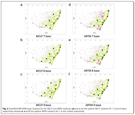
If you start with a clean slate and optimally position bases for either population coverage or average response time, both models place bases to cover that part of the coast (see Figure 2).  Hardly surprising.  When we modelled to optimise the existing base structure by adding or moving one or two bases, the mid north coast was either first or second location chosen by either model too.

Figure 2

This seems an obvious outcome from even a glance at the population distribution and current coverage in Figure 1.  What is surprising is that the 2012 review of the HEMS system in NSW (not publically released) which utilised the same census data in demand modelling did not come to the same conclusion when two previous reviews in the 1990s and 2000s had recommended just such a change.  Certainly the Reform plan for helicopter services which was released the following year did not make any changes or additions to base locations leaving this significant gap still uncovered.

Wagga Wagga was the other location identified for a HEMS base in the 2004 review.  Interestingly it is favoured as the first relocated base when the existing structure is optimised for average response time by moving Canberra to this location.  But a Wagga Wagga base also was not mentioned in the reform plan.

What about the green fields?

When the green field modelling was done it is clear that the current NSW system mostly closely resembles the model optimised for average response time, rather than coverage.  The Wollongong base really justifies its location on this basis as it contributes to a better overall average response time.  Its population coverage falls entirely within the overlapping circles of the Sydney and Canberra bases so it makes no contribution here, at least if a 45min response time is used as the standard.

There was another aspect that interested us compared with Norway.  In Norway all aircraft have the same capability and this is also true for the recently tendered services in NSW.  The unusual feature in NSW though (unique to Australia although common in Europe in particular) is a dedicated urban prehospital service operating from a base near to the demographic centre of the largest population density – Sydney.  The performance characteristics of this service have been well described (by us, because I’m talking about the CareFlight service which I think does serve a useful function) previously and when it was operating with its own dispatch system was the fastest service of its kind in the world to our knowledge.

Like the Wollongong service it operates entirely within the population coverage circles of other bases, but it makes an enormous contribution to average response time.  When this rapid response urban service is added to the network of large multirole helicopters in NSW the average response time across the entire state falls by more than 3.5mins because that service is able to access more than 70% of the state population within its catchment zone, and significantly faster than the multirole machines.

This modelling only takes into account the response time benefit of the specialisation afforded by such as service.  We have previously been able to demonstrate that the service is also much faster in almost every other aspect of care delivering patients to the major trauma services in Sydney only a few minutes slower than the road paramedic system but with much higher rates of intervention and ultimately passage through the ED to CT scan faster than either the road paramedic or multirole retrieval systems in NSW.  At least this was the case when it had its own specialised dispatch system but that is a story we have discussed previously too.

There are recurrent themes here.  The Rapid Response Helicopter service adds significantly to the response capability in NSW whether you model it using advanced mathematical techniques or whether look at the actual response data compared with the alternative models of care.  Indeed the real data is much stronger than the modelling.  It seems that at least in large population centres in Australia there is a role for European style HEMS in parallel with the more traditional multirole Australian HEMS models that service the great distances of rural and remote Australia.  Different options can work alongside one another to strengthen the whole system and better deliver stuff that is good for patients – timely responses when they really need them. The capability differences however need to be reflected in dispatch systems that maximise the benefits which come with specialisation rather than a one size fits all tasking model that takes no account of those significant differences.

Every version of the numbers I look at tell the same story.


Notes and References:

While this post covers a few ways of looking at a tricky sort of problem, there are lots of clever people out there with insights into how these things work. If you have ideas or examples from your own area, drop into the comments and help people learn.

Now, the paper that’s just been published is this one:

Garner AA, van den Berg PL. Locating helicopter emergency medical service bases to optimise population coverage versus average response times. BMC Emerg Med. 2017;17:31. 

The paper on optimal base locations in Norway is this one:

Røislien J, van den Berg PL, Lindner T, et al. Exploring optimal air ambulance base locations in Norway using advanced mathematical modelling. Injury Prevention. 2017;23:10-15.

And if you like any of the posts on here, then maybe share them around. Or sign up for an update when new posts hit with the email sign on thing.


The Bind When It Comes to Using a Binder – Part 5

You might recall a series more than a bit ago from Dr Alan Garner covering lots of thoughts on pelvic fractures and what might make sense for prehospital care. Well, he’s back at it with a case to get things rolling.

It is amazing what you find when you go looking.

Those who are regular readers of the CareFlight Collective will be aware of my concerns about the use of pelvic binders in lateral compression (LC) type fractures.  You can find parts 1, 2, 3 and 4 here.  In short a binder in the context of a LC fracture replicates the force vector that caused the injury and may make fracture displacement worse.  There is evidence of this in both cadaver models and in real live trauma patients.  However blind use of binders without knowing the fracture type (and even where it is known to be LC) has been considered safe as there were no reports that patients had deteriorated after application – until now.

Last year one of our teams applied a binder to a haemodynamically stable patient with a LC fracture.  There was immediate haemodynamic deterioration and new leg length discrepancy which had not been present prior to application of the binder.  The case report has been accepted for publication by the Air Medical Journal and about now would be a good time to say thanks to our co-authors from Westmead Hospital, Jeremy Hsu and Anne Douglas.  You can find a copy of the accepted manuscript accepted manuscript here.  You need to go and have a read of the manuscript then come back for the following comments to make sense so I suggest you do that now.

Go on…


I can wait …


Continuing…

Now that you have read the case report you can appreciate that this incident caused us considerable angst.  We knew this was theoretically possible but it was still a shock when it actually happened.  It has caused us to review our practice around binders to try and find the safest approach.

But at the same time we need to acknowledge that we live in a space of considerable uncertainty because we don’t have radiographs to guide our management in prehospital care.  All we have is our reading of the mechanism (which is often pretty unclear), the clinical state of the patient and perhaps a finding of pubic symphysis diastasis on ultrasound to guide us.  We have to acknowledge that we are going to get this wrong a reasonable proportion of the time.

So here is our reasoning and the place we ended up.

Firstly we need to remember that there is still no study of any kind (RCT or cohort) that has shown a statistically significant improvement in survival with binders.  There is some suggestive case series data (mostly in anterior compression or “open book” fracture types) and the benefit observed is raised BP and possibly blood product usage, not survival.  That is it.  As it seems we can definitely cause harm, it is worth keeping in mind just how poor the evidence for benefit is as we work our way through the approach to binder application.  One of my very experienced colleagues refers to binders as “pelvic warmers” due to the almost complete lack of evidence of benefit and I can’t tell him he is wrong.

First…

The first thing to consider is the stability of the patient.  Placing binders in stable patients with a possible mechanism has been considered acceptable practice despite the theoretical risks and indeed it is the policy of our local Ambulance service in NSW to do exactly that.

Other services such as Queensland have a more conservative approach.  They position the binder if there is a suggestive mechanism but only tighten it if the patient is unstable or becomes so.  Given that there is absolutely zero evidence that haemorrhage has ever been prevented by placing a binder I think the Queensland approach is a good one.   I know that there are reports of binders reducing fractures so perfectly that they have been hard to identify on subsequent imaging and it is impossible to say whether they would have bled without the binder, but benefit from prophylactic use has not even been investigated let alone proven.  And since we have now demonstrated that you can take a stable patient and turn them into an unstable one the summary of the published evidence now is:

  • Harm from binder application in stable patients = 1
  • Benefit from binder application in stable patients = 0

I acknowledge that prevention of haemorrhage is fundamentally difficult to prove but we have decided to join the Queenslanders.  We will position it in stable patients if we are suspicious but it is only tightened if and when the patient becomes unstable.  First do no harm.  If they are haemodynamically stable you can’t make things better, but you can makes things a whole lot worse.

Second…

Our next consideration as per the previous posts parts 1-4 is the mechanism.  If it is clearly a lateral compression fracture then there is not even a biologically plausible way a binder can help.  If you are doing an interfacility transfer, you have an Xray and it is a LC fracture, do not apply a binder no matter how haemodynamically unstable the patient is.  Every reported case who has had a rise in BP associated with a binder has had either anteroposterior compression (the majority of cases) or a vertical shear injury.  Therefore the evidence base for lateral compression fracture so far is:

  • Harm from binder application in patients with LC injury = 1
  • Benefit from binder application in patients with LC injury = 0.

Just don’t do it.

Now of course prehospital it can be really hard to know what the fracture type is.  But there are occasions where it can only be a lateral compression such as in MVAs where the impact is directly into the patient’s door with intrusion against their pelvis laterally.  Here is an example repeated from part 3:

Crash copy


In this case the car has slid into the pole sideways.  The impact is directly into the driver’s door who has been pushed across the cabin partially onto the passenger seat breaking the centre console in the process.  This can only be a lateral compression fracture and that is indeed what was found on pelvic plain film in the ED.  We no longer put binders on these patients, no matter how unstable they are – the binder has no plausible mechanism by which it can improve things.

Third…

The last part of the equation for us was the policy of application by the local Ambulance service which I have already mentioned.  We often turn up to find that a binder has already been applied.  Should we take it off again if stable?  If unstable and it really looks like a lateral compression injury?  The damage if any has probably already been done.  We are operating in an evidence free zone here of course.  Our consensus of opinion was that if it was properly applied we should just leave it there.

So we derived an algorithm which works through these steps in the reverse order that I have discussed them as that is the workflow in the real world:

Binder Algorithm

So the only patients who get a binder placed and tightened are the unstable patients where lateral compression is not likely from what we can see of the mechanism or we just don’t know the mechanism.  If you re-read part 3 this is the group we are suggesting that ultrasound may help in the decision making.  Benefit (in terms of improved BP, not survival) has only been demonstrated in patients with a widened symphysis so perhaps this is your single best clue that you have identified a patient who is likely to benefit from the intervention – if such a group actually exists.

The Wrap

The belief that pelvic binders are a benign intervention is becoming widespread even though there are already reports of serious complications such as massive necrosis from pressure injury (have a look here).  No intervention helps all patients, and all interventions carry risk.  The key is identifying the patients where the benefit outweighs the risk.  Given that proof of benefit from binders does not yet exist, think very carefully about the risk that you could make things worse by tightening it and converting a stable patient into an unstable one.  Use it only where the possibility of benefit outweighs the risk and there is just no possibility of benefit in a known lateral compression injury.  It can therefore never be justified if you know that is the injury type.  Similarly there is zero evidence of any kind for prophylactic use in stable patients, just a theory and even the theory does not make sense in lateral compression.

I find it difficult to believe that this is the first time a patient has deteriorated with a binder – we are just the first group to report it because we have been looking.  Complications are typically poorly reported in prehospital care for a number of cultural reasons (see Davis’ classic work on prehospital intubation where significant complications were picked up only by examining the monitor output; it was not reported by the clinicians).  Perhaps the temporal relationship between the binder and deterioration is not as clear as in this case, or the patient is already unstable and it is not possible to differentiate the additional bleeding caused by the binder from the bleeding that was already happening.  Or the subsequent instability is not attributed to the binder by the caregivers who think “just as well we put the binder on” without realising they actually caused it.

We would be really interested to hear if anyone else has observed this too.  But you won’t notice if you don’t look.  In the meantime I think we all need to examine our practices to ensure that are only applying the devices where there is a possibility that the patient will benefit from this as yet unproven intervention.  If there is no possibility of benefit, just don’t do it.


Notes:

You could always start with public cases like this to reflect on what we could do differently with pelvic binders.

Here’s the thing on the pressure necrosis with a pelvic binder again:

Mason LW, Boyce DE, Pallister I.   Catastrophic myonecrosis following circumferential pelvic binding after massive crush injury: A case report doi:10.1016/j.injury.2009.01.101

And if you’re interested in the stuff on this site you can always find the spot on this page to get your email in there