Tag Archives: pelvic fractures

The Bind When It Comes to Using a Binder – Part 5

You might recall a series more than a bit ago from Dr Alan Garner covering lots of thoughts on pelvic fractures and what might make sense for prehospital care. Well, he’s back at it with a case to get things rolling.

It is amazing what you find when you go looking.

Those who are regular readers of the CareFlight Collective will be aware of my concerns about the use of pelvic binders in lateral compression (LC) type fractures.  You can find parts 1, 2, 3 and 4 here.  In short a binder in the context of a LC fracture replicates the force vector that caused the injury and may make fracture displacement worse.  There is evidence of this in both cadaver models and in real live trauma patients.  However blind use of binders without knowing the fracture type (and even where it is known to be LC) has been considered safe as there were no reports that patients had deteriorated after application – until now.

Last year one of our teams applied a binder to a haemodynamically stable patient with a LC fracture.  There was immediate haemodynamic deterioration and new leg length discrepancy which had not been present prior to application of the binder.  The case report has been accepted for publication by the Air Medical Journal and about now would be a good time to say thanks to our co-authors from Westmead Hospital, Jeremy Hsu and Anne Douglas.  You can find a copy of the accepted manuscript accepted manuscript here.  You need to go and have a read of the manuscript then come back for the following comments to make sense so I suggest you do that now.

Go on…


I can wait …


Continuing…

Now that you have read the case report you can appreciate that this incident caused us considerable angst.  We knew this was theoretically possible but it was still a shock when it actually happened.  It has caused us to review our practice around binders to try and find the safest approach.

But at the same time we need to acknowledge that we live in a space of considerable uncertainty because we don’t have radiographs to guide our management in prehospital care.  All we have is our reading of the mechanism (which is often pretty unclear), the clinical state of the patient and perhaps a finding of pubic symphysis diastasis on ultrasound to guide us.  We have to acknowledge that we are going to get this wrong a reasonable proportion of the time.

So here is our reasoning and the place we ended up.

Firstly we need to remember that there is still no study of any kind (RCT or cohort) that has shown a statistically significant improvement in survival with binders.  There is some suggestive case series data (mostly in anterior compression or “open book” fracture types) and the benefit observed is raised BP and possibly blood product usage, not survival.  That is it.  As it seems we can definitely cause harm, it is worth keeping in mind just how poor the evidence for benefit is as we work our way through the approach to binder application.  One of my very experienced colleagues refers to binders as “pelvic warmers” due to the almost complete lack of evidence of benefit and I can’t tell him he is wrong.

First…

The first thing to consider is the stability of the patient.  Placing binders in stable patients with a possible mechanism has been considered acceptable practice despite the theoretical risks and indeed it is the policy of our local Ambulance service in NSW to do exactly that.

Other services such as Queensland have a more conservative approach.  They position the binder if there is a suggestive mechanism but only tighten it if the patient is unstable or becomes so.  Given that there is absolutely zero evidence that haemorrhage has ever been prevented by placing a binder I think the Queensland approach is a good one.   I know that there are reports of binders reducing fractures so perfectly that they have been hard to identify on subsequent imaging and it is impossible to say whether they would have bled without the binder, but benefit from prophylactic use has not even been investigated let alone proven.  And since we have now demonstrated that you can take a stable patient and turn them into an unstable one the summary of the published evidence now is:

  • Harm from binder application in stable patients = 1
  • Benefit from binder application in stable patients = 0

I acknowledge that prevention of haemorrhage is fundamentally difficult to prove but we have decided to join the Queenslanders.  We will position it in stable patients if we are suspicious but it is only tightened if and when the patient becomes unstable.  First do no harm.  If they are haemodynamically stable you can’t make things better, but you can makes things a whole lot worse.

Second…

Our next consideration as per the previous posts parts 1-4 is the mechanism.  If it is clearly a lateral compression fracture then there is not even a biologically plausible way a binder can help.  If you are doing an interfacility transfer, you have an Xray and it is a LC fracture, do not apply a binder no matter how haemodynamically unstable the patient is.  Every reported case who has had a rise in BP associated with a binder has had either anteroposterior compression (the majority of cases) or a vertical shear injury.  Therefore the evidence base for lateral compression fracture so far is:

  • Harm from binder application in patients with LC injury = 1
  • Benefit from binder application in patients with LC injury = 0.

Just don’t do it.

Now of course prehospital it can be really hard to know what the fracture type is.  But there are occasions where it can only be a lateral compression such as in MVAs where the impact is directly into the patient’s door with intrusion against their pelvis laterally.  Here is an example repeated from part 3:

Crash copy


In this case the car has slid into the pole sideways.  The impact is directly into the driver’s door who has been pushed across the cabin partially onto the passenger seat breaking the centre console in the process.  This can only be a lateral compression fracture and that is indeed what was found on pelvic plain film in the ED.  We no longer put binders on these patients, no matter how unstable they are – the binder has no plausible mechanism by which it can improve things.

Third…

The last part of the equation for us was the policy of application by the local Ambulance service which I have already mentioned.  We often turn up to find that a binder has already been applied.  Should we take it off again if stable?  If unstable and it really looks like a lateral compression injury?  The damage if any has probably already been done.  We are operating in an evidence free zone here of course.  Our consensus of opinion was that if it was properly applied we should just leave it there.

So we derived an algorithm which works through these steps in the reverse order that I have discussed them as that is the workflow in the real world:

Binder Algorithm

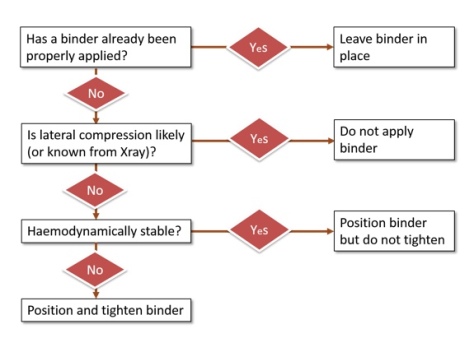
So the only patients who get a binder placed and tightened are the unstable patients where lateral compression is not likely from what we can see of the mechanism or we just don’t know the mechanism.  If you re-read part 3 this is the group we are suggesting that ultrasound may help in the decision making.  Benefit (in terms of improved BP, not survival) has only been demonstrated in patients with a widened symphysis so perhaps this is your single best clue that you have identified a patient who is likely to benefit from the intervention – if such a group actually exists.

The Wrap

The belief that pelvic binders are a benign intervention is becoming widespread even though there are already reports of serious complications such as massive necrosis from pressure injury (have a look here).  No intervention helps all patients, and all interventions carry risk.  The key is identifying the patients where the benefit outweighs the risk.  Given that proof of benefit from binders does not yet exist, think very carefully about the risk that you could make things worse by tightening it and converting a stable patient into an unstable one.  Use it only where the possibility of benefit outweighs the risk and there is just no possibility of benefit in a known lateral compression injury.  It can therefore never be justified if you know that is the injury type.  Similarly there is zero evidence of any kind for prophylactic use in stable patients, just a theory and even the theory does not make sense in lateral compression.

I find it difficult to believe that this is the first time a patient has deteriorated with a binder – we are just the first group to report it because we have been looking.  Complications are typically poorly reported in prehospital care for a number of cultural reasons (see Davis’ classic work on prehospital intubation where significant complications were picked up only by examining the monitor output; it was not reported by the clinicians).  Perhaps the temporal relationship between the binder and deterioration is not as clear as in this case, or the patient is already unstable and it is not possible to differentiate the additional bleeding caused by the binder from the bleeding that was already happening.  Or the subsequent instability is not attributed to the binder by the caregivers who think “just as well we put the binder on” without realising they actually caused it.

We would be really interested to hear if anyone else has observed this too.  But you won’t notice if you don’t look.  In the meantime I think we all need to examine our practices to ensure that are only applying the devices where there is a possibility that the patient will benefit from this as yet unproven intervention.  If there is no possibility of benefit, just don’t do it.


Notes:

You could always start with public cases like this to reflect on what we could do differently with pelvic binders.

Here’s the thing on the pressure necrosis with a pelvic binder again:

Mason LW, Boyce DE, Pallister I.   Catastrophic myonecrosis following circumferential pelvic binding after massive crush injury: A case report doi:10.1016/j.injury.2009.01.101

And if you’re interested in the stuff on this site you can always find the spot on this page to get your email in there

The Bind About Pelvic Binders – Part 4

Is this the last bit for now? Dr Alan Garner following up on pelvic binders after all the stimulating comments. If you haven’t already, check out part 1, part 2 and part 3.

During the writing of part three of this series on pelvic fractures and particularly after reading Julian Cooper’s comments (thank you Julian) I realised that the observational data around pelvic binders does not entirely fit with the theories. Let’s start with the theory and I might directly borrow Julian’s comments from Part 2 as he says it better than I could:

“In any type of pelvic injury. the bleeding will be either:

  1. Venous or bone ends: in which case keeping things still with a binder is likely to allow clot formation (low pressure bleeding, low or high flow).
  2. “Slow” arterial (the sort of thing seen as a blush on contrast CT) which will probably trickle on even with a binder but at a rate which is compatible with survival to hospital and (ideally) interventional radiology if they don’t stabilise spontaneously (high pressure, low flow bleeding).
  3. “Fast” arterial (e.g. free iliac rupture) which is likely to be fatal whatever one does, binder or not (high pressure, high flow bleeding).”

I need to state right up front that I agree with all of this. It all seems entirely reasonable and there is some cadaver evidence that movement of fractures associated with patient movement (e.g. sliding a patient from stretcher to a bed) is reduced when a binder is applied. It seems reasonable that a binder might slow, or at least reduce aggravation of venous and bone end bleeding with movement. It might even help the “slow” arterial bleeders too.

So what is my issue with all this? Studies like the Tan paper (15 patients) describe a dramatic and immediate increase in blood pressure associated with applying a binder to an “open book” style fracture and reducing it. Mean arterial pressure increased from 65mmHg to 81 and HR fell from 107 to 94 per min 2 minutes after application. The effect was associated with (although of course not necessarily caused by) reduction of the fracture. Nunn’s series of 7 patients showed even more dramatic changes in blood pressure measured at 15 minutes post binder application although they do not report the degree of fracture reduction achieved. Again we are dealing with tiny numbers of patients but the effect seems consistent – in shocked patients with anteroposterior compression or mixed type injuries who have a binder applied the blood pressure usually immediately rises (note one patient in Tan series who significantly deteriorated). In Nunn’s series with BP reported at 15 mins post application it is possible that the pelvis was “stabilised” and then a big fluid bolus was given but this cannot be the case in the Tan series where the effect is seen immediately.

Stabilising the pelvis against further movement and stopping venous and bone end bleeding cannot be the mechanism for this sudden rise in BP. Even stopping the “slow” arterial bleeders could not create such an immediate effect.

So what is going on? Warning – brainstorming not supported by any evidence following:

  • Compression of arteries in the pelvis resulting in increased systemic vascular resistance? (warned you about the brain storming – this seems pretty unlikely to me)
  • Compression of distended venous spaces causing a fluid shift back into the central circulation and increased BP. If this is the case then what you are seeing is a MAST suit effect and this has been shown to not necessarily be a good thing if you don’t also stop the bleeding.
  • One of my colleagues suggested it is pain associated with binder application that is causing the BP rise? Again doubt this is the case. Also not sure this is helpful if you are not also stopping the bleeding (as per MAST suit issues)

I don’t actually have a good theory for what is going on here but the effect is very clearly described in the literature. It seems to be a good thing although the Nunn paper in particular notes that ongoing volume resuscitation and other measures to stop the bleeding are usually then required. If anyone has any theories on what is happening here then please share with the rest of us.

A Recap

I might summarise the literature on pelvic binders as:

  • No study has yet demonstrated a significant decrease in mortality associated with binders
  • Increased fragment displacement, haemodynamic deterioration and some really ugly pressure injures (have a look at the case report by Mason for an absolute shocker) have been described with their use i.e. they are not benign.
  • They might decrease venous and bone end bleeding by preventing movement but we currently have no direct evidence to support this. Agree that this seems reasonable though.
  • An improvement in haemodynamics is often seen immediately at the time of application of a binder in shocked patients with an open pubic symphysis. Mechanism for this is currently unknown and we don’t have enough evidence to know whether this is actually a good thing or not. Going right back to part 1 of this series we should be very cautious about using surrogates such as improved BP as measures of outcome or binders may turn out to be MAST suit Mark 2.

I don’t want to be a wet blanket but I do believe that this is a realistic evaluation of the current evidence.

The Bottom Line on What I Do

Do I personally use binders prehospital?

Yes I do unless the injury is clearly lateral compression. I also am not afraid to loosen it again if the patient deteriorates. I think they are helpful for the open symphysis patients based on the documented haemodynamic improvement often seen in these patients but I acknowledge that I am hoping that this BP rise translates into lower mortality but I don’t have evidence to support this. I definitely will never criticise someone who has not put one on as there is just not enough evidence one way or the other.

Time for a segue – and perhaps a paradigm shift.

Come this way for other new thoughts but no more bad visual puns, people of the future. [Via Alan Kotok on flickr under CC 2.0]
The Ones Who Need More

Let’s look at Julian’s group 3: – ”Fast” arterial (e.g. free iliac rupture) which is likely to be fatal whatever one does, binder or not (high pressure, high flow bleeding). Again I agree with Julian here. These patients can die in minutes as is usually the case if you lacerate a vessel the size of the iliac artery, and there is absolutely nothing you can do about it prehospital.

Or is there?

Another thing I was taught as a boy is that if you can’t control arterial bleeding at the haemorrhage site then get proximal control. So how can you get proximal control for a punctured iliac artery? Clearly we are talking about occluding the aorta here but how do you achieve this prehospital?

The idea of REBOA (resuscitative endovascular balloon occlusion of the aorta) in the prehospital context has been getting a bit of attention with London HEMS recently introducing it. Now this sounds really sexy but it requires a skilled doctor with an ultrasound machine, time and good access to the patient. What I am proposing is the much simpler version of REBOA where the E stands for “External”.

Conflict of interest statement: Neither I nor either of my employers have a financial interest in the manufacture or distribution of the device I am about to mention – I just think it is a really cool idea.

AAJT copy

The device is the Abdominal Aortic and Junctional Tourniquet (AAJT) (here’s the link to the manufacturer’s website for their obviously positive coverage). A reasoned discussion on the relative merits of AAJT over traditional endovascular REBOA and some of the literature on both approaches can be found here.

The nice thing is that it sits around the waist and does not limit access to groins so that endovascular REBOA remains an option when you hit the trauma centre. If you can get one of these things on fast enough then even free rupture of an iliac vessel will potentially be controllable.

There are no reports yet of this device being used in catastrophic pelvic fracture haemorrhage but there are lots of reports of manual compression of the aorta being used in other causes of massive pelvic haemorrhage such as penetrating trauma, post partum haemorrhage and pelvic surgery. There are reports of the device being successfully used for massive bilateral lower limb injury in the military context. It should work in pelvic fracture too if proximal control is the key (famous last words).

The AAJT seems like the ideal prehospital device as you can place it in about 45 secs, in some situations you may be able to place it in a patient who is still trapped or whilst in transit to the hospital. That is just not going to happen with endovascular REBOA. And of course you don’t need a highly skilled physician with an ultrasound machine. Might have lower sex appeal factor but if occluding the aorta saves lives, this device is going to save far more lives than endovascular REBOA as it can be applied by a lot more people in a wider variety of situations. It is possible to put on an AAJT as well as a Pelvic binder as the binder sits around the greater trochanters and the AAJT is positioned over the umbilicus.

My own service has now acquired some AAJTs and we are about to introduce them to service. We will try and update you on our experience as it is early days yet for this device.

Lastly apologies to Julian if I have in any way misrepresented his opinions or taken his comments out of context. His comments certainly got me thinking however and that is what the Collective is supposed to be about so thanks Julian for contributing.

References:

Mason LW, Boyce DE, Pallister I.   Catastrophic myonecrosis following circumferential pelvic binding after massive crush injury: A case report doi:10.1016/j.injury.2009.01.101

The Bind About Pelvic Binders (Part 2)

This is part 2 in Dr Alan Garner’s series on pelvic fractures and the approach to binders. You can find part 1 here

In part one we had a look at the evidence for benefit from pelvic binders. In short there is no study yet published showing a significant improvement in mortality. Not even a cohort study.

Of course, it still might be OK to use them if they possibly help as long as there is no evidence of harm either (and they don’t cost too much). The probability of good has to outweigh the probability of evil. It is the potential for evil that I want to examine now so we can see where the balance lies.

Before we can do that though we need to have a quick look at the types of pelvic ring fractures (no one is suggesting that non-pelvic ring fractures of the pelvis benefit from a binder). So sorry folks but we have a bit of theory to re-visit.

Forces Down There

I use the Young and Burgess classification system as it is based on the force vector that caused the injury. In the prehospital world mechanism of injury is almost the only guide to injury type that is available to us (ultrasound may also give us some clues but we will talk about that in part 3).

AP compression injuries

AP Compression copy

This is an anteroposterior (AP) compression injury. This is the kind of fracture you see in frontal motor vehicle collisions, commonly in motor bike riders, and people who have been crushed by a vehicle rolling over their pelvis for example. The hallmark is pubic diastasis with or without disruption of the SI joints. The AP compression causes the pelvis to open: one or both hemipelves undergo external rotation.

External rotation of the hemipelvis results in an increase in the volume of the pelvic cavity which then allows more pelvic haemorrhage to occur before the osseous and soft-tissue structures cause tamponade. Exsanguination is the primary risk & reduction of the increased pelvic volume is one of the goals of prehospital care.

When I was a boy Master taught me the way to reduce a fracture is to reverse the force that caused it in the first place. With this type of injury a pelvic binder makes biomechanical sense because it reverses the direction of the force which caused it. In severe AP compression injuries one or both hemipelves have been rotated backward. Applying a binder will rotate the hemipelves back towards each other, or “close the book”.

Book copy

As I mentioned in part 1 there is very little evidence on whether this is actually helpful despite the theoretical benefit. Tan’s study was observational and involved only 15 subjects in an emergency department setting. All subjects had been X-rayed prior to application of the device so the type of injury was known (unlike our context in most cases). Nine of the 15 patients in this study had AP compression type injuries with wide diastasis of the pubic symphysis.   Although there is some missing data, all patients with this pattern either had no change in MAP or it improved. So far so good.

There is a similar English study with 3 severe AP compression injury patients who improved with a binder (Nunn) but numbers are obviously pretty small.

Croce’s study appears to have had mostly AP compression fracture types (186 patients with breakdown between types not stated). Decreased transfusion requirements were found in the binder group at 24 and 48 hours (significant), the patients had decreased length of stay (significant), and lower mortality (non-significant). This does provide some support for use in severe AP compression injuries noting the methodology issues which I discussed in Part 1 with a retrospective study that included patients over a 10 year period.

There are a number of other studies which show improved alignment +/- blood pressure rise in AP compression type fractures in trauma patients, in cadavers and even in one prehospital study. None of these studies assess patient outcome though (I acknowledge this is difficult in cadaver studies!) Reduction can be so good that the fracture is difficult to see on subsequent Xray.

So in AP compression injury all the evidence points to better anatomical alignment, higher blood pressure, lower transfusion requirements, and shorter length of hospital stay when you use a binder. Mortality might be better too, but this remains to be proven. The important thing is there are no reports of adverse events in this group. When you see this fracture type on Xray or the mechanism suggests this injury – go for the binder. The risk of adverse advents is certainly outweighed by the possible benefits based on the best current evidence.

Lateral compression injuries

Lateral compression copy

Lateral compression injury results in internal rotation of the affected hemipelvis. This internal rotation decreases rather than increases the pelvic volume so they tend to bleed less than the other types. Life threatening haemorrhage is still possible though. The hallmarks include sacral buckle fractures and horizontal pubic rami fractures.

Remember my boyhood teaching – “Grasshopper, to reduce fracture you must reverse force that caused it”. There is an obvious problem here as applying a binder replicates the causal force and if anything is likely to make it worse.

Have a look at this Xray of a lateral compression injury. Put a binder around the greater trochanters and pull. Are you a force for good or evil?

X-ray copy

So what is the evidence? The Tan paper did not include any lateral compression injuries – remember that they had looked at the X-ray prior to application. I assume they looked and thought “well that is not going to help”. There is no evidence the Croce study included any either.

Is there evidence that a lateral compression fracture can get worse with a binder? (You have to be suspicious when binder studies appear to have avoided this fracture type altogether).

A recent Australian study (Toth) from 2012 had 8 cases with lateral compression that had binders applied. In three it resulted in increased pelvic deformity on subsequent Xray. They did not report the haemodynamic consequences. In the other 5 there was no improvement. There is biomechanical evidence of this in cadavers too e.g. Bottlang et al (if you look at this paper note again that they did not even attempt it in the LC3 injuries – the most severe grade).

Now this really disturbs me. There are docs I have met who are adamant that pelvic fracture patients should not be logrolled & should only be moved on scoop stretchers etc because the fracture fragments might move just with this limited motion. These same docs are however happy to put a binder on regardless of mechanism and pull, creating a much larger force than a logroll does, when we have direct evidence that binders increase fragment displacement in lateral compression injuries. Some consistency would be nice.

The bottom line is that there is no theoretical reason to believe that binders help in lateral compression injuries and lots of reasons to think they might make things worse. There is direct evidence in real world trauma patients that increased deformity of the pelvis does occur. There is no published data at all on the haemodynamic consequences when this happens, but I am betting you are not going to see improvement. The balance of risk here is on the dark side, not the light.

Bottom line is leave the binder in the bag in the bag for clear lateral compression mechanisms. It cannot help and there is published evidence of harm.


(Stay tuned for part 3 where we’ll get to vertical shear injuries – and other stuff).

References:

Croce MA, Magnotti LJ, Savage SA, Wood 2nd GW, Fabian TC. Emergent pelvic fixation in patients with exsanguinating pelvic fractures. Journal of the American College of Surgeons 2007;204:935–9. [discussion 40–2]

Tan ECTH, et al. Effect of a new pelvic stabilizer (T-POD1) on reduction of pelvic volume and haemodynamic stability in unstable pelvic fractures. Injury (2010), doi:10.1016/j.injury.2010.03.013

Nunn T, Cosker TDA, Bose D, Pallister I. Immediate application of improvised pelvic binder as first step in extended resuscitation from life-threatening hypovolaemic shock in conscious patients with unstable pelvic injuries. Injury, Int. J. Care Injured (2007) 38, 125—128.

Bottlang M, KriegJ C, Mohr M, Simpson TS, Madey SM. Emergent management of pelvic ring fractures with use of circumferential compression. J Bone Joint Surg Am 2002;84-A(Suppl 2):43–7.