All posts by careflightcollective

A Bit About Paeds Trauma for Those Who Do A Bit of Trauma

This is a post put together by Dr Andrew Weatherall as background preparation for a talk at the SPANZA Paeds Update from March 14, 2015. This is an update for the occasional paeds anaesthetist. It’s not about covering it all but hopefully there’s a few useful points in there to prompt a little thought and discussion.

For lots of people who do a bit of paediatric care, there’s a bit of nervousness around little people. It’s a bit disproportionate to the numbers of actual cases of course because paeds trauma is not common. In fact, rates are slowly going down.

There is also a common paediatric conundrum to deal with – what do you do with adult evidence? This is because overwhelmingly trauma literature deals in the bigger, smellier version of Homo sapiens.

So the challenge is to provide a refresher on something that is getting less common for most of us, using evidence for other patients.

This might be easier with a story, weaved from a bit of experience and not that much imagination.

Crash copy

The Call Comes In

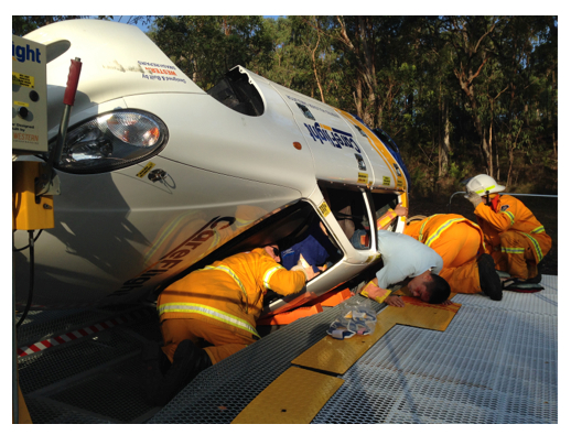
You get a call from the emergency department that they are expecting a paediatric patient from a crash, not too far from your hospital out on the far edges of the city. The road speed limit is around 80 km/hr and they have a 6 year old child who was sitting in the rear right passenger seat, in a booster seat. He’s probably too small for this booster seat. It doesn’t look like he was well secured.

The child was initially GCS 12/15, with a heart rate of 145/min, BP 85/58, a sore right upper quadrant, and a deformed right upper leg. Initially SpO2 was 96% but is now 100% on oxygen.

Where Should They Go?

Of the schools of thought (big kids’ centre vs place where they do lots of trauma but not lots of kids), NSW has gone for the hospitals with the pretty waiting rooms.

Probably the most relevant local research on the topic is from Mitchell et al. who looked at trends in kids going to paeds trauma centres or elsewhere. They found kids getting definitive care at a paeds trauma centre had a survival advantage 3-6 times higher those treated at an adult trauma centre.

There are issues with this. Mortality as a sole marker when you’re only discussing about 80 kids across 6 years may not be the most reliable marker of quality care. You only need one or two cases to shift from one column to the other to significantly skew the picture.

Possibly the more significant finding was the delay created by making that one stop. Stopping at another hospital (even within the metropolitan area) delayed arrival at the paediatric trauma centre by 4.4-6.3 hours. Early discussions to transfer obviously need to become a priority.

In NSW, the policy is now for ambulance officers to go directly to the paeds trauma centre if it’s possible within 60 minutes. Unless they don’t think they’ll get there.

The impact on the doctor working outside the kids trauma centres is two-fold:

  • There’s less paeds trauma to see.
  • The paeds trauma you do see will be the bad stuff.

Great mix.

The room with the international colour coding of "kids bay"
The room with the international colour coding of “kids bay”

At Emergency

So the patient, let’s call him  Joe, arrives. For the sake of discussion I’m going to assume he did come to the paeds trauma centre, but there’s a whole separate (possibly more interesting) scenario you could think through where he goes to a smaller metropolitan hospital.

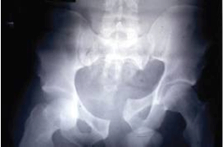
Joe arrives with an IV cannula in place and Hartmann’s running. He has a hard cervical collar in place. His GCS has improved to 14/15 (he’s closing his eyes but he seems a little scared) but his heart rate is now 155/min and his BP is 78/50. Peripheral oxygen saturations are still 100% on oxygen (they were 96% off oxygen). He is sore and tender in his right upper quadrant just like they promised. That right femur does look broken. There’s also a lump on the right side of his head, towards the front just on the edge of the hairline.

The New Alphabet

We all remember the alphabet, whether  first drummed in by the fluffy denizens of Sesame Street, or mostly embedded by a trauma course. A then B then C.

Anyone working in trauma knows this is only the older version. So 1900s. The trauma alphabet now has a bunch of variations (C-A-B-C,  MH-A-B-C, choose your edit) to highlight the need to think about arresting blood loss early.

A lot of this shift in thinking is surely related to the vast amount of knowledge gained in managing trauma from military conflict where stopping haemorrhage is one of the most effective things you can do to save lives.

The causes may be different (especially in kids), but some of the thinking can be transferred.

This makes sense not just because bleeding is not great for patients. It’s also because many of the measures required to stop it take more than a couple of minutes. Not so much in the case of tourniquets or fancy dressings that make you clot. Things like surgery, or interventional radiology, or blood product management.

If you’re an occasional paediatric trauma practitioner, there’s a few points worth remembering if you’re going to elevate the importance of haemorrhage control, even while getting the other stuff done:

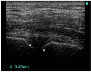
  • Find the blood early – better rapid diagnostic options, particularly ultrasound, need to be deployed early to figure out where blood loss might be happening.
  • Decisions need to support stopping bleeding – if the patient is bleeding, it is more than a bit important to progress continually towards making them not bleed. This is particularly relevant to arranging radiology and surgery as quickly as possible where indicated.
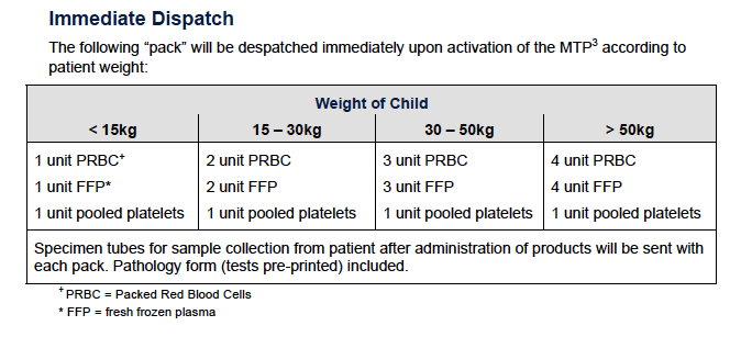
  • Transfusion – bleeding patients don’t need salty fluids. They need blood. And given what we know about acute traumatic coagulopathy, they probably need it in a ratio approaching 1:1:1 (red stuff: plasma:platelets).
  • Give TXA – after CRASH-2 and MATTERs, tranexamic acid has also made it to kids. A fuller discussion is over here (and there’s also the Royal College of Paediatrics and Child Health thing here though as I mention in that other post, I think they’ve got the doses not quite right).
Set 1 from The Children's Hospital at Westmead Massive Transfusion protocol (obviously, check local policies).
Set 1 from The Children’s Hospital at Westmead Massive Transfusion protocol (obviously, check local policies).


And here's the next delivery pack. (And check it out in full context, don't just rely on this screengrab.)
And here’s the next delivery pack. (And check it out in full context, don’t just rely on this screengrab.)

Joe is Getting Better

Ultrasound confirms some free fluid in the abdomen. The fractured femur is reasonably well aligned but you’ve started warmed blood products early. Joe is responding to his first 10 mL/kg of products with his heart rate already down to 135/min and a BP of 88/50. Respiratory status is stable. GCS is 15/15 and you’ve supplemented his prehospital intranasal fentanyl with IV morphine. 

You decide to go to the CT scanner to figure out exactly what is going on with the abdominal injury. Once around there Joe vomits and starts to get agitated. CT confirms a right front-temporal extradural haematoma. As he’s deteriorating you head up to theatres. 

photo 2

Now I’m going to assume anyone reading this is pretty happy with an approach to rapid sequence induction with in-line stabilisation to manage spinal precautions (not that we’d have a hard collar anyway, because those are on the way out in the draft ILCOR guidelines). We’d all agree on the need for ongoing resuscitation. I’ll also assume no one is going to stop the surgeons from fixing the actual problem while you mess about getting invasive arterial blood pressure measurement and a central line sorted.

What would be nice is some better evidence on what are the right blood pressure targets.

What BP target for traumatic brain injury?

Still, the best the literature can offer is a bit of a ¯\_(ツ)_/¯

If you look at this review from 2012 the suggestions amount to:

  • Don’t let systemic mean arterial pressure go below normal for age.
  • It might be even better to aim for a systolic blood pressure above the 75th percentile.
  • If you do have intracranial pressure monitoring and can therefore calculate cerebral perfusion pressure, then aim for > 50 mmHg in 6-17 year olds and > 40 mmHg in kids younger than that.

Hard to escape the thought we need more research on this.

The Rest of Joe’s Story

Everyone performs magnificently. Joe’s extradural is drained. His femur is later fixed and his intra-abdominal injuries are managed conservatively. The next most important thing might just be that you remembered to give him good analgesia.

Not Forgetting the Good Stuff

I might have some professional bias here, but I think remembering analgesia is just as important as the rest of it. Studies like this one suggest surprisingly high rates of PTSD symptoms even 18 months after relatively minor injury (38% though it was a small study). Although the contributors to PTSD are complex there is some evidence (certainly in burns patients)  that early use of opioid analgesia is associated with lower rates of PTSD symptoms.

This stuff matters. A kid with PTSD symptoms is more than just an anxious kid. They are the kid who is struggling with school, struggling with social skills and generally struggling with the rest of the life they were supposed to be getting on with. Pain relief matters.

So it is worth prioritising good analgesia:

  • Record pain scores as a vital part of the record.
  • Block everything that is relevant (no child with a femur fracture should have an opportunity for a femoral block of some description missed).
  • Remember treatment as analgesia (don’t just leave the fracture like you found it, for example).
  • Give rapidly acting,titratable drugs as a priority with regular checks of efficacy.
    • For example, fentanyl 5 mcg/kg in a 10 mL syringe gives you 0.5 mcg/kg/dose if you give 1 mL at a time. Do this and reassess every 3 minutes.
    • Likewise, ketamine 1 mg/kg in 10 mL provides a dose of 0.1 mg/kg each time you give 1 mL (though some would say you should use midazolam to offset dysphoria too).
    • Don’t forget novel options – methoxyflurane anyone?

The Wrap

Paeds trauma may not be as common, but it needs to be done to the same high standards we expect of trauma care anywhere. Most of the stories in resuscitation are well worn tales. But there are a few things to really take away:

* Think about doing everything to stop bleeding early.

* More blood for resuscitation, but more sensibly too.

* Never forget pain relief.


And with any luck, most of this is already old news.


Postscript: Just after I put this together, the always excellent St Emlyn’s blog put up something covering the latest changes to APLS teaching. To my immense relief a lot of it is the same. It’s worth checking out.

After the postscript: This isn’t designed to be too prescriptive and everything should be figured out in local context. Obviously any thoughts anyone has to share would be very welcome. 

A Bit of New Evidence on Drowning

An opportunity for a quick post to point to a new publication with something useful on drowning. From Dr Alan Garner.

Unfortunately we attend a number of paediatric drownings in the Sydney area every year. Many recover well. Some do not. Some do unexpectedly well. We have had a patient who was GCS 3 at our arrival and asystolic on the monitor make a full recovery. Most children in this situation however either die or are severely impaired.

This brings us to a vital question – when is it reasonable to stop resuscitation? Well, here’s some evidence to help inform the chat.

The Dutch Study

Over at the BMJ a new paper has just hit the screen:

Outcome after resuscitation beyond 30 minutes in drowned children with cardiac arrest and hypothermia: Dutch nationwide retrospective cohort study. 

This study is a nationwide observational study in the Netherlands of children with cardiac arrest due to drowning. The authors have put together ten years of data collected in a country with more than 30 million people. It seems unlikely we’ll see a bigger study.

The study indicates that no child resuscitated for more than 30 minutes had a good outcome. There were good outcomes in those resuscitated for less than 30 minutes.

This matches our experience. Our patient with the GCS 3/asystole combination and a subsequent good outcome had a return to spontaneous circulation while still on scene.

The other point of interest is that it is from an environment where water temperature is presumably a fair bit colder than the coastal fringes of Australia, but the results would appear to be similar.

It would appear that discontinuing resuscitation after 30 minutes in those with no neurological improvement or stuck in asystole is a reasonable practice.

It still comes down to time.


The Bind About Pelvic Binders – Part 4

Is this the last bit for now? Dr Alan Garner following up on pelvic binders after all the stimulating comments. If you haven’t already, check out part 1, part 2 and part 3.

During the writing of part three of this series on pelvic fractures and particularly after reading Julian Cooper’s comments (thank you Julian) I realised that the observational data around pelvic binders does not entirely fit with the theories. Let’s start with the theory and I might directly borrow Julian’s comments from Part 2 as he says it better than I could:

“In any type of pelvic injury. the bleeding will be either:

  1. Venous or bone ends: in which case keeping things still with a binder is likely to allow clot formation (low pressure bleeding, low or high flow).
  2. “Slow” arterial (the sort of thing seen as a blush on contrast CT) which will probably trickle on even with a binder but at a rate which is compatible with survival to hospital and (ideally) interventional radiology if they don’t stabilise spontaneously (high pressure, low flow bleeding).
  3. “Fast” arterial (e.g. free iliac rupture) which is likely to be fatal whatever one does, binder or not (high pressure, high flow bleeding).”

I need to state right up front that I agree with all of this. It all seems entirely reasonable and there is some cadaver evidence that movement of fractures associated with patient movement (e.g. sliding a patient from stretcher to a bed) is reduced when a binder is applied. It seems reasonable that a binder might slow, or at least reduce aggravation of venous and bone end bleeding with movement. It might even help the “slow” arterial bleeders too.

So what is my issue with all this? Studies like the Tan paper (15 patients) describe a dramatic and immediate increase in blood pressure associated with applying a binder to an “open book” style fracture and reducing it. Mean arterial pressure increased from 65mmHg to 81 and HR fell from 107 to 94 per min 2 minutes after application. The effect was associated with (although of course not necessarily caused by) reduction of the fracture. Nunn’s series of 7 patients showed even more dramatic changes in blood pressure measured at 15 minutes post binder application although they do not report the degree of fracture reduction achieved. Again we are dealing with tiny numbers of patients but the effect seems consistent – in shocked patients with anteroposterior compression or mixed type injuries who have a binder applied the blood pressure usually immediately rises (note one patient in Tan series who significantly deteriorated). In Nunn’s series with BP reported at 15 mins post application it is possible that the pelvis was “stabilised” and then a big fluid bolus was given but this cannot be the case in the Tan series where the effect is seen immediately.

Stabilising the pelvis against further movement and stopping venous and bone end bleeding cannot be the mechanism for this sudden rise in BP. Even stopping the “slow” arterial bleeders could not create such an immediate effect.

So what is going on? Warning – brainstorming not supported by any evidence following:

  • Compression of arteries in the pelvis resulting in increased systemic vascular resistance? (warned you about the brain storming – this seems pretty unlikely to me)
  • Compression of distended venous spaces causing a fluid shift back into the central circulation and increased BP. If this is the case then what you are seeing is a MAST suit effect and this has been shown to not necessarily be a good thing if you don’t also stop the bleeding.
  • One of my colleagues suggested it is pain associated with binder application that is causing the BP rise? Again doubt this is the case. Also not sure this is helpful if you are not also stopping the bleeding (as per MAST suit issues)

I don’t actually have a good theory for what is going on here but the effect is very clearly described in the literature. It seems to be a good thing although the Nunn paper in particular notes that ongoing volume resuscitation and other measures to stop the bleeding are usually then required. If anyone has any theories on what is happening here then please share with the rest of us.

A Recap

I might summarise the literature on pelvic binders as:

  • No study has yet demonstrated a significant decrease in mortality associated with binders
  • Increased fragment displacement, haemodynamic deterioration and some really ugly pressure injures (have a look at the case report by Mason for an absolute shocker) have been described with their use i.e. they are not benign.
  • They might decrease venous and bone end bleeding by preventing movement but we currently have no direct evidence to support this. Agree that this seems reasonable though.
  • An improvement in haemodynamics is often seen immediately at the time of application of a binder in shocked patients with an open pubic symphysis. Mechanism for this is currently unknown and we don’t have enough evidence to know whether this is actually a good thing or not. Going right back to part 1 of this series we should be very cautious about using surrogates such as improved BP as measures of outcome or binders may turn out to be MAST suit Mark 2.

I don’t want to be a wet blanket but I do believe that this is a realistic evaluation of the current evidence.

The Bottom Line on What I Do

Do I personally use binders prehospital?

Yes I do unless the injury is clearly lateral compression. I also am not afraid to loosen it again if the patient deteriorates. I think they are helpful for the open symphysis patients based on the documented haemodynamic improvement often seen in these patients but I acknowledge that I am hoping that this BP rise translates into lower mortality but I don’t have evidence to support this. I definitely will never criticise someone who has not put one on as there is just not enough evidence one way or the other.

Time for a segue – and perhaps a paradigm shift.

Come this way for other new thoughts but no more bad visual puns, people of the future. [Via Alan Kotok on flickr under CC 2.0]
The Ones Who Need More

Let’s look at Julian’s group 3: – ”Fast” arterial (e.g. free iliac rupture) which is likely to be fatal whatever one does, binder or not (high pressure, high flow bleeding). Again I agree with Julian here. These patients can die in minutes as is usually the case if you lacerate a vessel the size of the iliac artery, and there is absolutely nothing you can do about it prehospital.

Or is there?

Another thing I was taught as a boy is that if you can’t control arterial bleeding at the haemorrhage site then get proximal control. So how can you get proximal control for a punctured iliac artery? Clearly we are talking about occluding the aorta here but how do you achieve this prehospital?

The idea of REBOA (resuscitative endovascular balloon occlusion of the aorta) in the prehospital context has been getting a bit of attention with London HEMS recently introducing it. Now this sounds really sexy but it requires a skilled doctor with an ultrasound machine, time and good access to the patient. What I am proposing is the much simpler version of REBOA where the E stands for “External”.

Conflict of interest statement: Neither I nor either of my employers have a financial interest in the manufacture or distribution of the device I am about to mention – I just think it is a really cool idea.

AAJT copy

The device is the Abdominal Aortic and Junctional Tourniquet (AAJT) (here’s the link to the manufacturer’s website for their obviously positive coverage). A reasoned discussion on the relative merits of AAJT over traditional endovascular REBOA and some of the literature on both approaches can be found here.

The nice thing is that it sits around the waist and does not limit access to groins so that endovascular REBOA remains an option when you hit the trauma centre. If you can get one of these things on fast enough then even free rupture of an iliac vessel will potentially be controllable.

There are no reports yet of this device being used in catastrophic pelvic fracture haemorrhage but there are lots of reports of manual compression of the aorta being used in other causes of massive pelvic haemorrhage such as penetrating trauma, post partum haemorrhage and pelvic surgery. There are reports of the device being successfully used for massive bilateral lower limb injury in the military context. It should work in pelvic fracture too if proximal control is the key (famous last words).

The AAJT seems like the ideal prehospital device as you can place it in about 45 secs, in some situations you may be able to place it in a patient who is still trapped or whilst in transit to the hospital. That is just not going to happen with endovascular REBOA. And of course you don’t need a highly skilled physician with an ultrasound machine. Might have lower sex appeal factor but if occluding the aorta saves lives, this device is going to save far more lives than endovascular REBOA as it can be applied by a lot more people in a wider variety of situations. It is possible to put on an AAJT as well as a Pelvic binder as the binder sits around the greater trochanters and the AAJT is positioned over the umbilicus.

My own service has now acquired some AAJTs and we are about to introduce them to service. We will try and update you on our experience as it is early days yet for this device.

Lastly apologies to Julian if I have in any way misrepresented his opinions or taken his comments out of context. His comments certainly got me thinking however and that is what the Collective is supposed to be about so thanks Julian for contributing.

References:

Mason LW, Boyce DE, Pallister I.   Catastrophic myonecrosis following circumferential pelvic binding after massive crush injury: A case report doi:10.1016/j.injury.2009.01.101

Revisiting Old Stories About Little Airways

Dr Andrew Weatherall returns to stuff about paediatric airways, a bit of a companion to an earlier post with some practical tips. 

There are some things you’re taught from a very young age to believe in. Then it turns out it’s just plain wrong. Santa Claus. The Tooth Fairy. The Public Holiday Numbat. (Well, the last one might be specific to my upbringing.)

And in medicine there are plenty of examples those too. Oxygen is always good. You can’t manage trauma without a cervical collar. Then of course there’s pretty much everything about the paediatric airway. As if managing kids didn’t come with challenges anyway, we all get to work with information that is just plain wrong.

And there’s no mistaking that clinicians find paediatric airways difficult. The staff from Royal Children’s Hospital Melbourne have recently published a sizeable prospective study of emergency department intubations. This is from a big, clinically excellent tertiary kids’ hospital receiving 82000 patients in their ED every year. In 71 intubations across a year (only 71!), 39% had adverse events (most commonly hypotension in 21% and desaturation in 14%) and the first pass success rate was 78% (only 49% had a first pass intubation with no complications).

Now lots of things will contribute to those figures. But at least part of pondering that has to be making sure we understand what we’re dealing with.

"Please, go on" says Public Holiday Numbat [unchanged via quollism on flickr under CC]
“Please, go on” says Public Holiday Numbat [unchanged via quollism on flickr under CC]

Old Truths

Some old historical truths are harder to pull away than a spider web stuck to a bear with superglue. There’s a recent review that appeared in Pediatric Anesthesia written by Dr J Tobias  which steps through some of this dogma.

It points out that some of the classic teaching on the paediatric airway come out of a 1951 report by a Dr Eckenhoff. This includes the issues of the position of the larynx, the shape of the epiglottis and the funnel-shaped airway. Actually, to really trace the story, you have to start a little earlier.

Stepping Back

It’s 1897. Waistcoats aren’t ironic yet. Pipes aren’t an affectation they’re an expectation. Jack the Ripper is part of shared memory, not fevered historical narratives. And Bayeux was making casts of the airways of dead children. 15 casts actually in kids aged 4 months to 14 years.

Taking measurements of the circumference of the airway at the glottis, cricoid level and trachea, the cricoid ring was noted to be narrower than other parts of the airway (the topic of the shape of the airway wasn’t mentioned). This is the work that led to the idea that kids under the age of 8 had a conical larynx, with the cricoid ring as the narrowest point.

Consider for a second the qualities of plaster poured into a distensible tube. Wait, it’s not entirely distensible because the cricoid can’t distend. Is it maybe possible that the plaster may have distorted the anatomy? I’ll leave that with you for a bit.

This suggestion of the conical airway made its way into Eckenhoff’s later paper (though with a specific note that cadavers may not represent the living accurately). There were also some descriptive points raised:

  • The larynx moves down from the C3-4 level in the neonate to C4-5 in the adult (I’ve always been under the impression this move is brought about both by the need to phonate properly for speech and the loss of the need to breathe and breastfeed at the same time, but this point doesn’t feature in airway descriptions and I’m happy to be corrected).
  • A stiffer and more “U” or “V”-shaped epiglottis with an angle to the anterior pharyngeal wall of around 45 0 rather than lying close to the base of the tongue.
  • A case report of a 2 year old with airway complications thought to be related to an inappropriately sized tube, feeding the idea of uncuffed endotracheal tubes in kids under the age of 8.

All these points that form part of so much teaching lead to another question – would such a descriptive effort get a run in modern publishing?


Newer Tools Means Better Understanding

The answer of course is probably not. Of course you can only use what you have and it’s absurd to judge Eckenhoff (or Bayeux) for their accuracy against modern modalities. All we can do is revisit our thinking when new information becomes available.

We now have the significant advantage of radiological techniques (CT or MRI) and bronchoscopy to evaluate airways in children who aren’t dead. Again the Tobias article goes into more details but there are some key things to take from this modern literature:

  • In spontaneously breathing and muscle relaxed patients, the cricoid was not the narrowest part of the airway. That honour belongs to the vocal cords.
  • There is no change in the ratios of the cross sections over age – the cricoid doesn’t start relatively smaller and enlarge by the time you hit 8.
  • The cross-section looks like an ellipse (there’s more distance between the anterior and posterior bits than the two side bits).

 What should we do then?

Well for starters we should probably settle the tube choice thing. This is just more support for the argument to use a cuffed tube. For starters, the old “leak” test seems pretty dubious when you could be snug against the lateral walls but still leaking around the anterior or posterior areas. And I’m guessing no one has had their “leak accuracy assessment” externally audited.

It makes more sense to use an appropriately sized cuffed tube with the cuff pressure kept < 20 cm H2O. There’s now fairly convincing evidence that appropriately used cuffed tubes don’t cause big issues in recovery. Better ventilation, better monitoring, less flows and gentler tube material in contact with the mucosal wall. Makes sense.

What you can’t do is ignore the cricoid. It is still an unyielding bit of the anatomy and anyone can turn a high volume-low pressure cuff into a high volume-high pressure cuff – the difference is a couple of mL. And swelling in an airway that starts with a much smaller cross-sectional airway still means less margin for flow obstruction.

So choose the right tube, use it safely and you can get on with things.


While We’re At It, Let’s Forget One Blade to Rule Them All

Seeing as we’re talking about things that aren’t things, you may have also come across the idea that you should use a straight blade for the smaller kids (say, kids under 2). I’ve mentioned elsewhere that I think this is baloney but here’s a little bit of evidence.

Varghese and Kundu have published something on exactly this issue. 120 kids aged from 1-24 months had laryngoscopy (once anaesthetised and given muscle relaxation) with either a Miller or Macintosh blade, and then crossed over to the other type of blade. (Note they used both with the tip in the vallecula.)

The findings? The views were pretty much the same. The rates of difficulty were about the same. In fact, it’s a pretty beige set of numbers where being beige is actually as cool as things could be.

Some where the view wasn’t so great with a Macintosh had a better view with the Miller blade. Some went in reverse. The message though is a pretty resounding “same, same”.


So there’s just some truths that needed revisiting. There are no funnel-shaped airways. The airway isn’t round. There’s not one correct blade for the under 2s.

I still resent having to give up on the Public Holiday Numbat though.

References:

Here are the PubMed links for those mentioned in this post.

Long E, Sabato S, Babl FE. Endotracheal intubation in the pediatric emergency department. Pediatric Anesthesia 2014;24:1204-11. doi: 10.1111/pan.12490

Tobias JD. Pediatric airway anatomy may not be what we thought: implications for clinical practice and the use of cuffed endotracheal tubes. Pediatric Anesthesia 2015;25:9-19. doi: 10.1111/pan.12528

Varghese E and Kundu R. Does the Miller blade truly provide a better laryngoscopic view and intubating conditions than the Macintosh blade in small children? Pediatric Anesthesia 2014;24:825-9. doi: 10.1111/pan.12394

Why? How? What? Big Questions for Prehospital Simulation

At CareFlight another round of training many people is about to come up so it seemed like a good chance to ask Dr Sam Bendall for her first contribution. 

Sam is an Emergency Physician who is passionate about education, particularly all things simulation. She works half-time at Royal Prince Alfred Hospital in Sydney in the Emergency Department where she helped develop and teaches the RPA Trauma Team Training program, teaches on the CIN nurses programs and helped develop the ED Essentials program. 

At CareFlight she is a retrieval doc (the other half-time) and the Deputy Director of Education. The CareFlight Education Team are always up to interesting things – from training the Australian Defence Force medical on how to look after all things ballistic, medical, surgical and paediatric, to running the Pre-Hospital Trauma Course both in Sydney and other locations (Malaysia, anyone?), to running Trauma Care Workshops all over the country. Oh, and of course all those working with CareFlight too. 

Anyway, here’s Sam …


As a passionate advocate for simulation I look around and see this amazing tool appear in many guises, all under the same blanket term. It certainly seems to mean many things to many people depending on their previous experiences. In some ways it is the SWISS ARMY KNIFE   of kinesthetic education. The coolest knife has pliers and scissors. However, just like a Swiss army knife, it can be a harmful weapon (hence the name!), just plain useless (like when you want the one with pliers but you only have the single blade), or a bit uninspiring and encourage automatic behaviours – e.g. all Swiss army knives are red and you should have one.

This clearly rubbish version doesn't even have the magnifying glass.
This clearly rubbish version doesn’t even have the magnifying glass.

Simulation has almost become the learning apparatus du jour – everyone has to do it but some are not sure why or how to really make it work. A bit like having a Swiss army knife so you can be part of the Swiss army, but it lives in the drawer.

I will put a disclaimer in at this point. The following are my own opinions – the musings of a dedicated simulation-phile after several years of training in simulation and doing simulation exercises for anywhere from 2 – 150 people.

So what’s the point?

WHW copy                                            

I’m going to put a slightly different spin on it, with an emphasis on simulation for the pre-hospital environment. Simon Sinek, in his TED talk in 2009 titled “Start with Why” made a very powerful case for asking yourself WHY you want to do something… in this case, simulation, at the outset. The HOW and the WHAT will follow if you drill down onto the why and firmly establish WHY you want your participants to do simulation.

Simulation is a journey, for both the instructors and participants. Hopefully a journey towards some constructive learning, but one that will have many interesting twists and turns along the way. Being sure of WHY you are undertaking this part of the journey, gives you the freedom to explore the twists and turns of the journey without losing sight of the original intent. So my step 1 in building a simulation, is to ask yourself why? WHY are you doing this?

In our organisation, our WHY? is to create a mission-ready workforce.

Pre-hospital medicine throws out so many variables – communication, teamwork, environmental, situational awareness, medical challenges, geographical challenges and the list goes on.

In order to make our workforce mission ready, we need them to be critical thinkers, able to choose the right skill, equipment and approach for the right case at the right time.

Though if we had one of those shapeshifting Terminator ones could we program it to be friendlier?

We also need them to be aware of the variables they will need to deal with on real jobs so that they can manage them consciously. In order to do this we need to replicate as many of these variables as possible so they can address them in a training environment. We aim to send our participants out on jobs that feel just like the scenarios they have done in training. No pressure!

HOW? – choose your weapon

Weapon copy

The simulation menu is fairly extensive and limited only by your creativity and ability to structure it in a way that is true to the learning objectives and easy to follow for your participants. The key elements of creating a scenario, whether it be for 2 or 50 participants, is that they need to know the rules, boundaries, and premises for the scenario….. hmm sounds like parenthood!

So first decide on your structure. Is it an audience that is learning a concept for the first time and you need to do it for real, but slow it down? Well “pause and discuss” is your man. Do you need to see where your participants’ critical decision making is at and where the deficits lie? Immersive, relatively high fidelity simulation, with key variables built in, is the tool of choice.

Do you need to occupy 30 participants in a large scale simulation? – Create foci so the participants will need to form their own teams within the simulation. This will bring out all of the teamwork, communication and leadership points from the start.

Whatever weapon you choose, it needs to be appropriate to the audience, their experience and what you are trying to teach by doing the simulation exercise.

WHAT …the final frontier

Well this depends on what you are trying to deliver in your simulation. If, for example, your aim is to test and consolidate a new protocol, then the scale of your simulation can be quite limited. You may not need to bring in as many variables, or much fidelity. As long as the key prompts are there for the participants and they have the knowledge, skills and equipment to fulfill the protocol, then a limited scenario is fine.

BUT…. and there is a BIG but in this one. Be realistic in developing your scenario. If you are testing an ALS protocol, doing a bog standard ALS protocol with a patient in a bed may tick your box. BUT ….. in 20 years of medicine I think I have been to less than 10 arrests in ward beds and way more than 30 in other places – the toilet, the CT scanner, theatre, the foyer of the hospital, the waiting room, the beach etc. etc. You get the picture. So I would argue here that a bog standard ALS type scripted scenario has its place, but should be followed up by the application of the protocol where it is likely to happen and bring in the teamwork and communication aspects that we know actually make ALS protocols work in real life.

At CareFlight we educate using a “crawl, walk, run” paradigm.

  • First you crawl – i.e. you learn the skill or concept in isolation.
  • Then you walk – using relatively low fidelity simulation with limited learning outcomes, you learn to apply that skill appropriately.
  • Then you RUN. In our “RUN” scenarios, we introduce many more variables that replicate the environment they will have to operate in. We increase the fidelity and prompt the participants to evaluate the situation, decide whether that skill or concept is appropriate, apply it if it is or find an alternative if it is not. This layering approach helps to consolidate skills and knowledge and develops critical decision-making processes in a way that is directly applicable to the job we do.

When you get to the RUN scenario you are trying to bring out multiple learning points across many categories, for example:

  • Teamwork and communication (CRM)
  • Leadership skills (CRM)
  • Graded assertiveness and conflict resolution (CRM)
  • Scene safety and situational awareness (CRM, environmental and logistics)
  • Management of a multitrauma patient in an isolated environment (medical)
  • Packaging and preparation for transport (logistics, medical)
  • How to carry a patient out of the bush safely (logistics, medical)
  • Planning for contingencies e.g. weather etc. (logistics)

Then the scenario has to be much higher fidelity and be crafted in a way that replicates those key learning objectives – CRM, medical, environmental and logistic. You need to recreate the key environmental elements that will impact on the participants’ decision-making, bring in the key teamwork elements, replicate the equipment or types of equipment they will use and think about the team structures they will be given. Even simple tweaks to the scenario such as limiting access to the patient’s head, can improve the problem solving and CRM elements of the scenario so the devil is very much in the detail here. AND SO IS THE FUN …

Why yes that is a mobile rollover simulator that some clever people built ...
Why yes that is a mobile rollover simulator that some clever people built …

A Short Video About Bleeding Airways

Managing the airway in prehospital and retrieval medicine is a challenge and has inspired many a discussion in many a setting. And anyone working in the area would appreciate the additional challenge when there’s lots of blood getting in the way.  As a result everyone has tips and and tricks to try and manage things.

This is by no means the first time people have come up with an approach (or shared an approach) but in the spirit of wide-ranging discussion, here’s a suggestion from Dr Alan Garner recorded for posterity in video.

It runs for about 10 minutes and you’ll note that at the end there’s an update as the approach evolved.

All thoughts, feedback and experience very welcome.

Keeping Things Calm: Remote Retrieval of the Psychiatric Patient

Jodie Mills, RN works with CareFlight’s Top End Medical Retrieval Service, flying out of Darwin across vast stretches of the Northern Territory. She grew up in the Royal Melbourne Hospital ICU before moving to Darwin 8 years ago where she completed midwifery studies.  She joined CareFlight 4 years ago and slightly pities all those who don’t get to fly in the top end. 


When asked to contribute to a blog and write about psychiatric aeromedical retrieval all I heard was my colleagues’ collective signs of “not another psych job!!”

The thing is, I’ve developed a bit of an interest in these patients after closely looking at the psychiatric retrievals in NT for the last 3 years. This specialised patient group presents a huge challenge to both the flight crew and our remote colleagues when presenting acutely unwell in our communities.

By the Numbers

I recently presented at the ASA/FNA/ASAM Aeromedical Retrieval Conference in Brisbane. I thought maybe we had a few psychiatric patients but I quickly realised after my presentation that the number of psychiatric retrievals we undertake in the top end is well above average i.e. its extremely high (15% of our total missions).

From Feb 2012 to the 20th October 2014 we retrieved 651 psychiatric patients, averaging 22-24 per month  – it’s an almost daily occurrence. Demographically the patient population remains consistent with approx. 90% of patients Indigenous Australians, with male to female ratio if 1.45:1. The mean age is 31, however our youngest was 12, our oldest being 74 years.

We have only intubated 3% of this population which has led to expedited admission to the singular psychiatric facility at Royal Darwin Hospital (RDH). The inpatient psychiatric ward at RDH has a catchment area of 700,000 square kilometres.

It’s Not Just a Local Thing

Mental illness throughout the world is on the increase with the WHO (2014) predicting mental illness to be second only to cardiovascular disease for burden of disease by 2030. The stigma associated with mental health issues remains the greatest obstacle to such patients accessing appropriate care. This stigma may be even more pronounced in remote Indigenous communities. Drug induced psychosis, predominantly cannabis (397 patients), followed by suicidal ideation/ hanging (224) were the most common diagnosis with the remaining patients having bipolar, mania or behavioural disturbances.

At the ASA conference I asked my aeromedical peers “How do you transport your psychiatric patients?” the answer was “we don’t, they go by road”. I quickly realised then that CareFlight and other retrieval services working in truly remote areas provide a unique service.

The small window view of a big country.

The Perfect Storm

We all know too well the challenges involved in the aeromedical transport of compliant patients who are unwell. However if we add delusions, hallucinations, physical aggression a tendency to physical violence and homicidal thoughts into the mix we have a potential aviation disaster on our hands. These are the just some of the symptoms the majority of our psychiatric patients display when referred to CareFlight. We then face the task of transporting such patients in a small aircraft where we will place seatbelts and wrist and ankle restraints on them, we will sit approximately 50cm away from them and the tell them they cannot smoke, they cannot go to the bathroom, they cannot eat or drink. I can’t imagine how stressful this must be for a patient that is already thought disordered.

What We Do

The biggest challenge for the aeromedical clinician is assessing the need and amount of sedation that will be required for safe retrieval of the acute psychiatric patient. If we have learnt anything it is definitely that “one-size DOES NOT fit all” when it comes to choosing sedative combinations to safely retrieve acute psychiatric patients. However we have found that pre-flight sedation with an atypical antipsychotic (olanzapine) and a sedative (diazepam) is of the utmost importance. As we become better skilled at treating psychiatric patients we have increased the pre-sedation (Olanzapine & Diazepam up to 20mg oral) which seems to be decreasing inflight sedation requirements. This enables the psychiatric patient to be admitted to the appropriate ward in a timely manner.

Top Tips for What to Do:

  1. Start sedation early:

As mentioned above, premedication prior to retrieval is vitally important. In most cases an antipsychotic (Olanzapine 10mg) and a benzodiazepine (Diazepam 10mg) is the premedication of choice. However, acute psychiatric patients presenting with drug induced psychosis (be it first or subsequent presentations) routinely require up to 20mg- 30mg of both Olanzapine and Diazepam orally. The first dose of sedation is given prior to the crew departing Darwin and then half an hour prior to the crews landing at the communities/ regional hospitals. This administration is overseen by the Medical Retrieval consultant (MRC) on duty. If the patient is not responding to the Olanzapine and Diazepam, the likelihood of requiring in-flight sedation is increased as is the probability of intubation for transport.

  1. In-Flight sedation:

We find in flight we tend to use midazolam, propofol and ketamine. The drug of choice is directly related to the flight doctor’s area of expertise. The ED Registrars tend to use midazolam and ketamine, whereas the ICU and Anaesthetic registrars head for the propofol and midazolam.

On arrival at the referral centre the patients are assessed for the need for further sedation prior to flight.

  1. Pre-Flight Sedation: Midazolam 2-5mg IV
  2. In-flight Sedation:
    • Propofol Infusion 0.2-0.5mg/kg/hr and titrate as required
    • Ketamine Infusion 0.5-1mg/kg/hr and titrate as required

A Richmond Agitation Sedation Scale (RASS) of -3 (Moderate) to -4 (Deep) or a Ramsey Sedation score of 5 indicates the level of sedation required for safe transport.

The ability to discontinue the sedative and allow the patient to wake prior to admission at the receiving centre is extremely important. If the flight crew are able to deliver an acute psychiatric patient to the receiving centre awake and ready for assessment this expedites the patients’ admission to the in-patient facility from the emergency department or, optimally allows for direct entry into the inpatient facility at the receiving centre.

Richmond Agitation Sedation Scale:                                                                               

Richmond copy

Ramsey Sedation Scale:

Ramsey copy

  1. Managing the environment:

Managing the stressors of flight is extremely important when retrieving an acute psychiatric patient. Using ear plugs, blankets to keep patients warm, positioning for comfort when heavily sedated, limiting cabin conversation and ensuring physical restraint are fastened appropriately ensures the acute psychiatric patient does not experience any extraneous stressors throughout their flight.

  1. Local law enforcement:

On occasion the local law enforcement will be involved with the acute psychiatric retrieval. The resource poor environment of the community clinic necessitates the presence of police to help control patients as documented under the section 9.

  1. Coordination:

The coordinating Medical Retrieval Consultant will liaise with the Consultant Psychiatrist on call at the hospital, alerting them to the impending admission.   The Consultant Psychiatrist then coordinates  with their in-patient team to ensure timely assessment of the patient if they are to be admitted through the emergency department.


Although the collective groan when another psychiatric retrieval arises resonates through the base we remain steadfast in our support to our rural and remote colleagues and we will continue to play a vital role in maintaining safety of the community, the families and the patients who are all touched by mental illness in the top end of the NT.


DIY to Stop the Blood

This thing comes from Dr Andrew Weatherall, paediatric anaesthetist and prehospital doc. He also blogs over at www.theflyingphd.wordpress.com


I don’t do DIY. This is partly because in the same way I wouldn’t expect a carpenter to have a crack at fixing their kids’ bones in preference to seeing an orthopod, I think it makes sense to use professionals.

It’s also because I’m just not that great at it. Anything I did make would end up looking like something trying to squeeze itself into the shape of the thing it is sort of supposed to be. And I’m fond enough of my family to want to protect them from the risks of my own handiwork.

Here's one I prepared earlier (via CC and flickr user mhlradio)
Here’s one I prepared earlier (via CC and flickr user mhlradio)

Anyway, I do paediatric anaesthesia. I get to spend more than enough time trying to make things that aren’t quite right for the situation fit in with what I need. Why DIY at home when you have to DIY at work?


Making Things Fit

The problem with paeds practice is that kids are sometimes kids and sometimes little adults and often forgotten in research. Or if not forgotten put in the category of “the ethics and logistics of that will be so painful I’d rather remove my spleen via my auditory canal”. And in trauma care we’re also dealing with total numbers that are lower than is the case for adults.

So what we end up with is lots of extrapolation from adult data and lots of retrospective studies sprinkled with the occasional fairy dust of a small case series. Then we have to try and mash those leftovers together to come up with a plan for a very specific situation.

An example: how about tranexamic acid in trauma?


Making It Up

Following on from CRASH-2 and MATTERs, what to do in the younger generation is an obvious question. A big prospective study in kids after trauma would be perfect. And a pipe dream.

So if you turn to the literature what you see is a large number of people trying out archery on summer camp and hitting many, many different targets while all shooting vaguely in the same area.

To corral some of them in one spot, take the review by Faraoni and Goobie looking at antifibrinolytics in non-cardiac surgery in kids. All of the following values are listed as loading doses in the scoliosis and craniofacial groups: 10, 15, 20, 50, 100 and 1000 mg/kg with infusions anywhere from 1 mg/kg/hr up to 100 mg/kg/hr. In the scoliosis patients there are total numbers of up to 80 patients and slightly baffling figures suggesting total blood loss is decreased but transfusion requirement pretty much the same. Or that in the craniofacial surgery group it seems like probably there might be slightly less blood loss and transfusion needs.

But in paediatric cardiac surgery there might be more seizures too, even though the overall safety profile looked pretty good. Nothing definitive though. Such clarity.

So now the job is to consider how to take this magnificently imperfect evidence and apply it to a specific and different clinical scenario, trauma.

Go.


The Pragmatist

The Royal College of Paeditrics and Child Health and the National Paediatric Pharmacists Group Joint Committee had exactly this challenge back in 2012. It’s the intellectual equivalent of trying to catch pancake batter. Messy.

Ultimately they chose what they termed the pragmatist’s option – 15 mg/kg loading (up to 1 g) over 10 minutes then an infusion of 2 mg/kg/hr. Maybe enough to do something, but with a homeopathic infusion so you were unlikely to get complications. Entirely rational in the absence of evidence too.

But what if there was another approach?


Another Way

What they didn’t have access to was some recent data out of the UK military Afghanistan experience in Camp Bastion. TXA had become standard for adult trauma patients under certain conditions after the release of CRASH-2 and both editions of MATTERs. These sort of treatment centres don’t just receive adults though and they must have been wrestling with what to do in smaller patients.

What they describe is another type of pragmatic approach. Rather than any adjustment they just did what they were already doing. Tranexamic acid in a 1 g dose for all comers and more on the basis of medical assessment (though it looks like no one got another dose).

This gets past lots of problems, particularly with getting accurate weights or ages and the need to learn different treatment regimes. It also comes with a certain amount of glee, not because you’re sort of saying “kids are just little adults” and you know that would break plenty of people. You’re actually saying “kids are adults”. If you say that 3 times while drawing a pentagram in a circle of candles, somewhere a paediatrician will be woken with a pain between their shoulder blades.

They describe a breakdown of 66 patients under 18 getting TXA and 700 without TXA. Having severe abdominal or extremity injuries and showing evidence of severe metabolic acidosis were significant predictors that TXA would be used. TXA use was independently associated with reduced mortality but no great difference in packed red blood cell/fresh frozen plasma transfusion ratios. Intriguingly in those getting a large volume transfusion, receiving TXA was associated with greatly improved neurologic status at the time of discharge (now that opens up a need for more work). They didn’t note an increased risk of thromboembolic complications (but they probably don’t have the numbers to be sure about that).

Overall, we’re talking about kids with an average age of 11 so using the equation of (3 x age) + 7, the weight might be about 40 kg (though I’m not certain if the weights might be a bit less than algorithms from developed countries). That would mean a starting dose averaging round 25 mg/kg.


The Other Extra Bit

That 2014 review also mentions an additional titbit that’s a little useful. Some pharmacokinetic work has been done in patients with craniofacial surgery patients and it appears that an upfront dose of 10 mg/kg then an infusion of 5 mg/kg/hr is optimal for establishing appropriate drug levels. This is far more useful information than cardiac surgery pharmacokinetics where additional considerations of dilution by bypass circuits, potential for pre-existing cyanosis and a variety of other factoids make it hard to draw comparisons. So 10 mg/kg might be enough initially but the subsequent infusion should probably be more than a scattering of holy water (as in more than 2 mg/kg).


The Bottom Line

We’re still stuck with not enough information about paediatric patients. Will there be a bigger study in paeds trauma soon? Probably not. But we can say with more confidence than before that doses that are pretty big seem to be OK.

So what would I do now? I’d modify the pragmatic plan and go with a 20 mg/kg loading dose (or 0.2 mL/kg of our current stock) and once in hospital I’d go with an infusion of 5-10 mg/kg/hr.

And I’d still hope someone is going to try to build a better shack.


References:

Are you after that review? It’s Faraoni D and Goobie SM. The Efficacy of Antifibrinolytic Drugs in Children Undergoing Noncardiac Surgery: A Systematic Review of the Literature. Anesth Analg 2014;118:628-36.  

Or maybe the RCPCH statement on using TXA in trauma – try here.

And here’s the Pubmed listing for the newer trauma study – Eckert MJ, Wertin TM, Tyner SD et al. Tranexamic acid administration to pediatric trauma patients in a combat setting: The pediatric trauma and tranexamic acid study (PED-TRAX). J Trauma Acute Care Surg. 2014;77:852-8. 

And in case you didn’t have it already, here’s the spot for the [(3 x age) + 7] calculation. Luscombe MD, Owens BD, Burke D. Weight estimation in paediatrics: a comparison of the APLS formula and the formula ‘Weight = 3(age)+7’ Emerg Med J 2011;28:590-3. 

Thoughts from the Control Tower

This is the first of what we hope will be a series of posts from Dr Paul Bailey who works as a Medical Director for CareFlight International Air Ambulance. Paul will try to provide insights into the challenges of managing retrievals across oceans. Here’s the starter. 

In his real life, Paul Bailey is an Emergency Physician based in Perth, Western Australia who dabbles in the Greyhound racing industry (having owned 10 dogs and never been to the track).  He can often be found in the outer at an Aussie Rules football oval, most commonly critiquing the performance of the umpires in an entirely constructive manner.  Past lives include a molecular biology PhD – in Jellyfish venom – don’t ask – and being a glassy in various drinking establishments in Western Australia. 

 Paul has previously undertaken international retrievals, helicopter work supporting Australian Army exercises and time with Queensland Rescue at Cairns. He now makes cameo appearances on the International Medical Director roster, as a medical director for retrievals in the NT and the Inpex oil and gas business.


If you’ve decided to be involved in retrieval, why think local when you can think global? After many years of toiling through school, then medical school, and then advanced training in the acute care specialty of your choice, you’re now in the hot seat ready to go.

Living the Dream

You’ve always fancied yourself as an airborne medico ever since you sat on your Dad’s knee watching “The Flying Doctors” in the late ‘80s and thinking how cool that would be. Truth be told, you also liked Top Gun and from time to time have drawn a laugh with the immortal line: “Negative Ghostrider, the pattern is full.” But you still buzzed the control tower anyway.

Fly Past copy

Or perhaps more likely you rode that little bit too close to your Mum on your BMX. You’ve also heard, along the way that everyone in retrieval gets a nickname and you’re tossing up between Maverick and Goose.

Top Gun copy

And it’s safe to say you have bought the Ray Ban Aviators already.


Living the Reality

So what’s it really like? Different to that, not surprisingly.

CareFlight International Air Ambulance (CFIAA) is an ever changing beast, with our clinical teams and aircraft based in Darwin and Sydney. Over the journey we have also had aircraft in Perth and Cairns. Depending on where the team is on duty, they are most likely to be flying between Indonesia, East Timor, Papua New Guinea, Darwin and Adelaide (for Darwin crews) and Fiji, Noumea, Norfolk Island, Sydney and Brisbane if you are based in Sydney. Of course, it’s international so there actually isn’t a spot on the globe that shouldn’t be thought of as up for grabs.

Co-ordinators and Medical Directors sit behind the team at all times and, due to the miracle of mobile phones and the internet can be almost anywhere. Many of these folk never meet in person but are always looking over everybody’s shoulder.

More of that later, in this awe inspiring opening to the series I though I’d start with how it all gets going – who pays for it all?


The People with the Deep Pockets – Travel Insurers and Governments

When your average citizen takes out travel insurance, it is most likely to protect against such tragedies as losing an iPhone overseas, dropping a wallet in the ocean or perhaps finding himself in Vietnam and his luggage in downtown Boston. Having been in this game a while now, it is my opinion that if losing your iPhone is the worst thing that happens on your holiday you’ve had a pretty good time.

Recliners copy

It may surprise you to know that a travel insurance policy is, by and large, a health insurance policy. Greater than 95% of the spend of travel insurance companies relates directly to health costs.

Each travel insurer has a series of service providers sitting behind them, one of which is an assistance company. There are a relatively small number of assistance companies that engage in this type of work. They have 24h call centres with co-ordinators, nurses and doctors (much like us).

In the event that John Q Citizen becomes unwell or is injured whilst they are away – by getting gored by a bull in Pamplona for instance – they or their relatives call the assistance company and a whole train of events unfolds, which might include directing patients towards local health care facilities to help with their medical problems.

The assistance company will maintain contact with the now patient and their family, and depending on how things in the event that the medical issue is of a serious nature, things tend to pan out in one of two ways that relate to the quality of health care available locally and the underlying urgency of the patient’s medical condition.

Let’s focus on the sicker end of the spectrum because clearly many issues are of a minor nature and never come anywhere near us.

If “definitive care” is available locally AND the quality of local medical care is high AND treatment of the matter is urgent assistance companies will usually head down the route of electing to keep the patient where they are for treatment in the theory that that is both (a) best for the patient and (b) cheaper than an international medical retrieval.

If “definitive care” is NOT available locally OR the quality of local medical care is questionable it is then the key decisions become urgency and mode of transport.  There are, again, an array of transport options available but seeing as we are in the Air Ambulance business, again we might focus on that.


Send in the Big Bird

The assistance company, having decided that medical evacuation is required and that this is most appropriately by air ambulance asks its panel of Air Ambulance providers for a quote. At this stage, the available information usually consists of the patient location and their ultimate destination. No clinical information is available.

Our co-ordinator submits a quote, and due to the price of aviation fuel the retrieval company with the aircraft that has to do the least amount of flying to get the job done is usually cheapest. Paperwork is exchanged and the job confirmed.

Clinical information is then available and at this stage the CFIAA Medical Director is brought into the discussion – to liaise with treating clinicians at the hospital of origin as well as the destination unit. How many times is it quick and easy to have a chat with someone you’ve never met in the hospital? Well, it is fair to say that these conversations can be difficult – finding the right person in an overseas hospital at a sometimes odd times of day and surmounting the language barrier is not straightforward.

We are often going to locations where the quality of the medical and nursing staff are excellent but the broad array of diagnostic equipment that many of us consider routine are just not available. Similarly there are many locations where the patient will have a problem that is unable to be treated effectively with the resources available locally. It’s part of the game, and in many ways it’s why we are needed in the first place. A lot of the legwork for the coordinator is about trying to construct a story that is useful for the retrieval team and help plan for every contingency.

So, that’s a summary of all the things that happen before you get to find out about a case. We haven’t even got to the challenges of the actual patient yet.

I might finish off with a thought for the day:

If you can open your packs blindfolded, upside down and in a thunder storm – and know where everything will be, you have satisfactorily completed orientation.


The Bind When It Comes to a Binder (Part 3)

There’s been a lot of stimulating discussion after parts 1 and 2 of this series from Dr Alan Garner (you can check those here and here). Here’s part 3. 

Thanks for sticking with the discussion so far. In part 2 we had a look at AP compression injuries and lateral compression injuries. Short summary is binders make sense and there is some observational evidence of benefit in AP compression injuries. However in lateral compression, binders make no biomechanical sense and there is definite evidence they increase fracture displacement both in cadavers and real live trauma patients.

The final group that we have not yet considered in the Young and Burgess classification is the vertical shear group. These patients are complex because the injuries are both horizontally and vertically unstable. You will see what I mean if you have a look at this Xray:

Pelvic Xray copy

Is putting a binder around the greater trochanters and pulling going to help? Will it produce anatomical realignment? I think you will agree that it is hard to know. In this case it might rotate the left hemipelvis inward and create even more distortion. You might also guess that some traction on the left leg before you apply the binder might get a better result too. More on this later.

Is there any actual evidence that things can get worse with a binder in vertical shear? Tan’s paper had six of this injury type. Two of the six had a fall in MAP immediately after the binder was applied, one by 20mmHg! It is a bit crazy that we are discussing studies with six patients but this is the level of published evidence to date. Such as it is, the evidence is that one in three vertical shear injuries deteriorated immediately after the binder was placed. Toth’s paper found that 14/17 patients had improved alignment post binder in this group so it often does some good. Unfortunately you have to think really carefully about this group, and be prepared to loosen it off again if you don’t get the response you were hoping for.

Yes, loosen it if the patient deteriorates! Primum non nocere. Remember there is as yet no study that has shown significant mortality improvement with pelvic binders. They are not a standard of care. If what you do makes thing worse then backing off is the right thing to do. I try not to let my own psychological need to do everything I can for the critically injured patient in front of me drive me to do things that might actually harm the patient. Sometimes less is more.

So what are we left to conclude?

  • AP compression – makes biomechanical sense and low level evidence binders help
  • Lateral compression – makes no biomechanical sense and real world evidence binders increase fracture displacement. Is “just holding it still” enough?
  • Vertical shear – a really difficult group; evidence of haemodynamic and anatomical improvement in the majority but clinically significant deterioration has also been documented
  • The real world as always is a bit more complex than this and mixed injury types happen
  • And of course, no evidence yet overall that binders have a significant effect on the outcome that matters in this case –mortality.

It should be pretty obvious that the type of fracture should be the guide to whether or not a binder might help. This is great if you are doing an interhospital transport and have an Xray. Not really helpful though if you are at the roadside, on an oil rig, or at a remote clinic 1000kms from the nearest trauma centre with no imaging (as our teams frequently are). So how can you work out whether a binder will help?

First thing is reading the injury mechanism. If you are at the scene you may get a lot of clues about the force vector, particularly in motor vehicle trauma. This is a photo of an incident I attended a few months ago.

Powerpole copy

In this case the car had slid sideways into the power pole striking the driver directly in the right side with such force that she had broken the centre console with the left side of her pelvis and was partially in the passenger seat. This can only be a lateral compression injury and there is no way a binder can help. Direct frontal injuries are also pretty obvious and the injury type is going to be an AP compression if a pelvic ring fracture is present.

This is good as far as it goes. It really does not help much with other mechanisms like pedestrians and motor cyclists. Were they side-on or front-on when they were hit by the truck? Motor cyclists can have a significant rotational component to their flight before they hit something which can make prediction of injury patterns really problematic.

There is one other trick which can give you a really valuable clue. Symphyseal diastasis is the hall mark of the AP compression injury. This is the sign that the “book is open”. If you can identify this then you can identify the group that is likely to benefit from a binder to “close the book” (although some will have vertical shear so care is still required). This is yet another use for my trusty companion, the handheld ultrasound.

The width of the pubic symphysis can easily be measured with the same high frequency linear probe that you use to exclude a pneumothorax. The upper limit of normal width measured at the point shown in the image is <25mm in adults (Bauman). I am not aware of any published data on children. As with all things there is a bit of variation here and cadaver studies have shown that anterior sacroiliac ligament disruption is likely for displacement greater than 45 mm and unlikely for values less than 18mm. So if the symphysis is less than 18mm you can be very confident the pelvis is not “open”.

Ultrasound copy

Clinical ultrasound copy

Note that in the source study for the reference range they failed to achieve a measurement in one case because the symphysis was wider than the width of their probe. You may have to move the probe from side to side to pick up both sides of a really wide symphysis.

If the patient does not have symphyseal widening on the other hand there is no reason to believe that a binder will help and they may well have an injury type that will be worsened by a binder (the symphysis does not open in lateral compression). Ultrasound is likely to be our best guide as to which patients have the possibility of benefitting from a binder whilst avoiding those where harm is the more likely outcome.   Some patients with vertical shear and an open symphysis may still deteriorate so there is no guarantee, but ultrasound will at least allow you to identify the group who have the possibility of benefit rather than harm.

As with so many things in prehospital care we need some good studies in this area. In the meantime, read the mechanism, read your ultrasound screen and be judicious in applying binders. Harm has occurred with these devices – they are not a universal panacea. Much of the art of medicine is picking the right patient for the intervention so you maximise benefit and minimise harm. This patient group is no different.

And thanks for the comments. Julian Cooper’s thoughts helped me work through my own theories on the issues and I have realised that our theories and the observational evidence don’t seem to align. There is also some potential new approaches to the massively haemorrhaging pelvis that are easily applicable in the prehospital environment and those are worth looking at too.

So looks like I am doing part 4. Stay tuned folks.

Bauman M et al. Ultrasonographic determination of pubic symphyseal widening in trauma: the FAST-PS study. The Journal of Emergency Medicine, Vol. 40, No. 5, pp. 528-–533, 2011.

Doro CJ et al. Does 2.5 cm of symphyseal widening differentiate anteroposterior compression I from anteroposterior compression II pelvic ring injuries? J Orthop Trauma. 2010 Oct;24(10):610-5. doi: 10.1097/BOT.0b013e3181cff42c.

Tan ECTH, et al. Effect of a new pelvic stabilizer (T-POD1) on reduction of pelvic volume and haemodynamic stability in unstable pelvic fractures. Injury (2010), doi:10.1016/j.injury.2010.03.013

Toth L, King KL, McGrath B, Balogh ZJ. Efficacy and safety of emergency non-invasive pelvic ring stabilisation. Injury, Int. J. Care Injured 43 (2012) 1330–1334