Category Archives: Prehospital Trauma

Sandpits, Better Eyes and New Monitors – Can NIRS work for prehospital medicine?

This is part 2 of a series (part 1 is here) on trying to study near-infrared spectroscopy in the prehospital setting by Dr Andrew Weatherall (@AndyDW_). Can NIRS work? No one can be sure but here’s one approach to getting some data we can actually use. 

A while back I did a post where I pointed out that when you get sold technology, there’s a whole history behind the machine that goes beep that means it’s probably not what you’re told. And the example I used was near-infrared spectroscopy tissue oximetry.

That was partly because I’m involved in research on NIRS monitoring and I’ve spent a lot of time looking at it.  Like every time I look carefully in the mirror, there’s a lot of blemishes that I miss on a casual glance. I also don’t mind pointing out those blemishes.

So that post was about all the things that could get in the way – light bouncing about like a pinball, humans being distressingly uncatlike, comparing monitors that are might be apples and aardvarks rather than apples and apples and basing your whole methodology on assumptions of tissue blood compartments. Oh, and maybe you can’t get sunlight near your red light.

Sheesh.

The thing is, I really want to answer that original question – “How’s the brain?”

So enough of the problems, can we find some solutions?

Actually I’m not certain. But I can say what we came up with. It’s a plan that involves sandpits, hiding numbers and finding better eyes. Oh, and changing the design of monitors forever and ever.


Playing in Sandpits

Our first step was to try and figure out if NIRS technology could even work in the places it wasn’t designed for. Not near the cosy bleeping of an operating theatre monitor where the roughest conditions might be inflicted by a rogue playlist.

We figured that the first issues might be all the practical things that stop monitors working so effectively. And we already knew that in the operating suite you often needed to provide shielding from external light to allow reliable measurements.

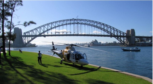
So we asked for volunteers, stuck sensors to their heads and took them driving in an ambulance or hopped on the helicopter to do some loops near Parramatta. It gave us lots of chances to figure out the practicalities of using an extra monitor too.

And we learnt a bit. That we could do it with some layers of shielding between the sensors and the outside world. That the device we tested, though comfortable next to an intensive care bed was a bit unwieldy at 6 kg and 30 cm long to be carried to the roadside. Most importantly that it was worth pushing on, rather than flattening everything in the sandpit and starting again.

Early engineering advice included "just put a tinfoil hat on everyone to shield the sensors". I just ... I ... can't ... [via eclipse_etc at Flickr 'The Commons']
Early engineering advice included “just put a tinfoil hat on everyone to shield the sensors”. I just … I … can’t … [via eclipse_etc at Flickr ‘The Commons’]

Hiding Numbers and Getting Out of the Way

The next thing that was pretty obvious was that we couldn’t set out to figure out what NIRS monitoring values were significant and at the same time deliver treatments on the basis of those numbers. We needed to prospectively look at the data from the monitor and see what associations were evident and establish which bits in the monitoring actually mattered for patients and clinicians.

Of course paramedics and doctors tend to like to fix stuff. Give them a  “regional saturation” number which looks a little like mixed venous oxygen saturation while the manufacturer (usually) puts a little line on the screen as the “good-bad” cutoff line is a pretty good way to see that fixing reflex kick in. So to make sure it really is a prospective observational study and we’re observing what happens in actual patients receiving their usual treatment we ended up with a monitor with none of the numbers displayed. Better not to be tempted.

It was also obvious that we couldn’t ask the treating team to look after the NIRS monitor because they’d immediately stop doing the same care they always do and occasionally (or always) they’ll be distracted by the patient from being as obsessive about the NIRS monitor as we need for research.

So recruiting needs a separate person just to manage the monitor. On the plus side this also means we can mark the electronic record accurately when treatments like anaesthesia, intubation and ventilation or transfusion happen (or indeed when the patient’s condition obviously changes). It’s all more data that might be useful.

Getting Better Eyes

One of the big problems with NIRS tissue oximetry so far seems to be that the “absolute oximetry” isn’t that absolute. When you see something claiming a specific number is the cutoff where things are all good or bad, you can throw a bucket of salt on that, not just a pinch.


Maybe this much salt. [via user pee vee at Flickr's 'The Commons']
Maybe all of this salt. [via user pee vee at Flickr’s ‘The Commons’]
The other thing is that to pick up evolving changes in a dynamic clinical environment is difficult. What if it isn’t just the change in oximetry number, but the rate of change that matters? What if it’s the rate of change in that number vs the change over the same time in total haemoglobin measurements, or balance between cerebral monitoring and peripheral monitoring at the same time? How does a clinician keep track of that?

What we might need is a way of analysing the information that looks for patterns in the biological signals or can look at trends. The good news is there’s people who can do that as it’s acutally a pretty common thing for clever biomedical engineers to consider. So there are some clever biomedical engineers who will be part of looking at the data we obtain. When they have spare time from building a bionic eye.

My bet is that if NIRS monitoring is ever to show real benefits to patients it won’t be only by looking at regional saturation (though we’ll try that too). It will be the way we look at the data that matters. Examining rapidly changing trends across different values might just be the key.


Thinking About the Monitors We Need

Let’s imagine it all works. Let’s assume that even with all those hurdles the analysis reveals ways to pretty reliably pick up haematomas are developing, or the brain is not receiving enough blood flow, or oedema is developing (and there are other settings where these things have been shown), there’s still a big problem. How do you make that information useful to a clinician who has a significant cognitive load while looking after a patient?

For each NIRS sensor that is on (3 in this study) we’ll be generating 4 measurements with trendlines. The patient is likely to have pulse oximetry, ECG, blood pressure and often end-tidal capnography too. Putting together multiple bits of information is an underappreciated skill that highly trained clinicians make a part of every day. But it adds a lot of work. How would you go with 12 more monitoring values on the screen?

Yes Sclater's lemur, that's 16 monitoring values to keep track of. [via user Tambako the Jaguar at flickr]
Yes Sclater’s lemur, that’s 16 monitoring values to keep track of. [via user Tambako the Jaguar at flickr]
So before we can take any useful stuff the analysis reveals and free clinicians to use the information, we need to figure out how to present it in a way that lets them glance at the monitor and understand it.

How should we do that? Well it’s a bit hard to know until we know what we need to display. My current guess is that it will involve getting clever graphics people to come up with a way to display the aggregated information through shapes and colours rather than our more familiar waveforms (and that’s not an entirely novel idea, other people have been on this for a bit).

So then we’d need to test the most effective way to show it before finally trying interventional studies.

This could take a bit.

And that is a story about the many, many steps for just one group trying to figure out if a particular monitor might work in the real world of prehospital medicine. There are others taking steps on their own path with similar goals and I’m sure they’ll all do it slightly differently.

I hope we end up bumping into each other somewhere along the road.


Notes and References:

Here’s the link to our first volunteer study (unfortunately Acta Anaesthesiologica Scandinavica has a paywall):

Weatherall A, Skowno J, Lansdown A, Lupton T, Garner A. Feasibility of cerebral near-infrared spectroscopy monitoring in the pre-hospital environment. Acta Anaes Scand 2012;56:172-7.

If you didn’t look on the way past, you should really check the video of Prof. Nigel Lovell introducing their version of a bionic eye. It’s pretty astonishing and I can’t quite believe I get to learn things from him.

It’s the very clever Dr Paul Middleton who was first author on a review of noninvasive monitoring in the emergency department that is well worth a read (alas, another paywall):

Middleton PM, Davies SR. Noninvasive hemodynamic monitoring in the emergency department. Curr Opin Crit Care. 2011;17:342-50.

Here’s the PubMed link from a team taking a preliminary looking tissue oxygen monitoring after out-of-hospital cardiac arrest:

Frisch A, Suffoletto BP, Frank R, Martin-Gill C, Menegazzi JJ. Potential utility of near-infrared spectroscopy in out-of-hospital cardiac arrest: an illustrative case series. Prehosp Emerg Care. 2012;16:564-70.


All the images here were via flickr’s ‘The Commons’ area and used without any modification under CC 2.0

Studies in Blood from Iran – A Quick Review

We all want to stop bleeding. Here’s a quick review from Dr Alan Garner of a paper coming out of Iran that looks at haemostatic dressings. 

Hatamabadi HR et al. Celox-Coated Gauze for the Treatment of Civilian Penetrating Trauma: A Randomized Clinical Trial. Trauma Monthly. 2014;20:e23862. dii: 10.5812/traumamon.23862

There is not a lot of data on haemostatic dressings in the civilian context and human data from the military context is not randomised for obvious reasons. It is therefore nice to see a RCT on this subject in humans. In the study they compare the time to haemorrhage control and amount of haemorrhage in stab wounds to the limbs between 80 patients treated with Celox gauze versus 80 patients treated with normal gauze.

The study is from an emergency department in Tehran and is pragmatic in design. There are some limitations of the study worth mentioning. It was open label, and the amount of bleeding was measured simply by the number of gauze squares used. Weighing the gauze would have been a more accurate way to estimate ongoing blood loss.

The details of how the gauze was applied isn’t that clear. To be effective the gauze needs to be packed into the wound against the bleeding vessel. Was the Celox used in this way to maximise the chances it would work? I can’t tell from the paper. Oh, and the company provided the product for the trial.

Perhaps the biggest puzzle in the design is that patients with really significant haemorrhage (those requiring transfusion) were excluded from the trial. This is the group where you really want to know if the stuff works. You could theorise that this group of patients may have trauma coagulopathy and the method of action of Celox (being by electrostatic attraction and independent of clotting factors) might be particularly useful and a bigger difference between groups may have been found. I guess that will have to wait for another day and another trial that someone works through ethics.

Acknowledging all of this, there was a significant difference in the time taken to achieve haemostasis and the amount of ongoing bleeding with the Celox gauze looked superior by both measures.

This suggests that it remains reasonable to use these products as evidence continues to point to efficacy. Of course these agents are not a magic bullet and all the other principles of haemostasis need to be applied as a package, including urgent transport to a surgical facility.

Examining the Hairs on the Yak – A Good Chance for More Chat

One of the good things about research that has its own issues, is that there is lots of scope to learn from the things about it that are good, as well as those that aren’t so great. The nice thing about ongoing comment is it gives even more chances to explain why a researcher might make certain choices along the way. Every question in research has more than one way of approaching some answers. Dr Alan Garner returns to provide even more background on this particular study, which has already generated some interesting conversation and a follow-up post

It’s an excellent thing to be able to keep having discussion around the challenges related to both conducting and interpreting a trial.  These things always bring up so many valuable questions, which deserve a response. So this is not going to be quick, but I hope you’ll have a read.

Lots of things changed between the time this trial was designed and now. Standards of care change. Systems, processes and governance models change. Indeed, in this trial standard care changed underneath us. We completed the protocol and gained ethical and scientific committee approval for this study during 2003.

The world was a different place then – at the start of 2003 George W Bush was US President and Saddam Hussein was still running Iraq. There is no keener instrument in medicine than the retrospectoscope particularly when focused 12 years back. Would I have done things differently if I knew then what I knew now – absolutely. Does the trial have hairs? Looks like a yak to me and I don’t think we are pretending otherwise.

Asking Questions

Did we ask the right question? The question was pragmatic. Add a doctor and with them comes a package of critical care interventions that were not routinely otherwise available in our standard EMS system. A number of cohort studies had previously looked at exactly this question and more studies have asked it since. Even papers published this month  have examined this question although the issue often overlaps with HEMS as that is how the doctors are frequently deployed.

I might segue slightly to address dp’s question as well which overlaps here. Is it the procedures that the team performs or the person performing the procedures that matter? Dp suggests that a better study design would be to have them all use the same protocols then we compare doctors with non-doctors. Such a randomised trial has actually been done although it is a long time ago now – 1987. It is one of the classic Baxt and Moody papers and was published in JAMA.

Patients were randomly assigned to a helicopter staffed by a flight nurse/paramedic or a flight nurse/emergency physician. The flight nurse and emergency physicians could perform the same procedures under the same protocols including intubation, chest tubes, surgical airways and pericardiocentesis. By TRISS methodology there was lower mortality in the group that included the doctor and the suggestion was this might be related to how they judged the necessity for intervention, rather than technical skill. This study is well worth a read. They note that the outcome difference might have been removed if the nurse/paramedic team was more highly trained but where does this end? We then move into the question of how much training is enough training and this is an area that I think is still in its infancy. Each time you do some research your prompt a whole lot of extra, usually interesting questions.

All That Methods Stuff

Anyway, back to this paper. All analyses presented in this paper were pre-specified in the statistical analysis plan. Although the protocol paper was not published till 2013, the statistical analysis plan (SAP) was finalised by the NHMRC Clinical Trials Centre in August 2010, more than a year prior to follow up of the last recruited patients. Copies of the SAP were then provided to the trial funders and NSW Ambulance at the time it was finalised in 2010. Along the way we have presented data in other settings, mostly at the request of interested parties (such as the Motor Accidents Authority who specifically requested analyses of road trauma cases) and in retrieval reviews. This is why there has been the opportunity for extra public scrutiny by experts like Belinda Gabbe. And public scrutiny is a good thing.

And Standard Treatments?

I’m very happy to provide some reassurance that this study did not rely on junior doctors being put through EMST/ATLS and then sent out to manage severe prehospital trauma patients. Rather the trial protocol states that treatment was according to ATLS principles. In 2003 there was no other external standard of care that we could cite for trauma patient management that was widely and internationally recognised.

The specialists had of course all completed EMST/ATLS but they were also all critical care specialists in active practice in major trauma centres in Sydney with ongoing exposure to severe trauma patients. The average years of prehospital trauma management experience held by this group of doctors at the beginning of the trial was more than 12 years each. They operated to those high level of treatment standards, with regular reviews of management to make sure this was current best practice over the life of a trial that ended up being longer than we hoped.

Other Dimensions of Time

And time wasn’t a friend. Recruitment was indeed slower than planned. This is a common problem in prospective trials. Our estimates of how long it would take to recruit the required sample size were based on a written survey of the major trauma centres in Sydney in 2003 to determine how many unconscious adult blunt trauma patients they were seeing each year. This was reduced to 60% to reflect the fact the trial would recruit for only 12 hours each day (although during the busiest part of the day) and the time needed to recruit was then estimated at 3 years. We in fact planned for 4 years to allow for the fact that patients usually disappear when you go looking for them prospectively. This of course is exactly what happened but to a greater degree than we planned.

I agree it would have been nice to have the results formally published earlier. We did present some results at the ICEM in Dublin in June 2012. It is interesting to note that Lars Wik spoke immediately before me at this conference presenting the results of the CIRC trial on the Autopulse device. This study was finally published online in Resuscitation in March 2014, more than three years from recruitment of their last patient and this trial did not include a six month neurological assessment as HIRT did.   Getting RCTs published takes time. Given we did have to perform six month outcome assessments I don’t think we were too far out of the ball park.

Quokka copy 2
To keep you going, here’s a quokka who looks like he’d be up for a chat too. [Via Craig Siczak and unchanged under Creative Commons.]

Randomising in Time Critical Systems

Just to be sure that I really have the right end of the stick on the question of excluding patients after randomisation I ascended the methodology mountain to consult the guru. For those that don’t know Val Gebski he is Professor and Director, Biostatistics and Research Methodology at the NH&MRC Clinical Trials Centre in Sydney. He was our methodology expert from the beginning of planning for the trial.

When I reached the mountain top I had to leave a voice message but Val did eventually get back to me. He tells me excluding patients post randomisation is completely legit as long as they are not excluded on the basis of either treatment received or their outcome. This is why he put it in the study design.

These are essentially patients that you would have excluded prior to randomisation had you been able to assess them properly and of course in our study context that was not possible. The CIRC study that I have already discussed also adopted this approach and excluded patients that did not meet inclusion criteria after enrolment.

Prehospital studies where you have to allocate patients before you have been able to properly assess them are always going to have these kind of difficulties. The alternative for a prehospital RCT would be to wait until you know every element of history that might make you exclude a patient. How many of us have that sort of detail even when we arrive at the hospital?

Extra Details to Help Along the Discussion

The newly met reader might also like to know that the call off rate was about 45% during the trial, not 75%. This is not different to many European systems. If you don’t have a reasonably high call off rate then you will arrive late for many severely injured patients.

And of course the HIRT study didn’t involve “self-tasking”. The system randomised cases on a strict set of dispatch guidelines, not on the feelings of the team on the day. This process was followed for nearly 6 years. There was not a single safety report of even a minor nature during that time. Compliance with the tasking guidelines was audited and found to be very high. Such protocolised tasking isn’t inherently dangerous and I’m not aware of any evidence suggesting it is.

It’s reassuring to know that other systems essentially do the same thing though perhaps with different logistics. For example in London HEMS a member of the clinical crew rotates into the central control room and tasks the helicopter using an agreed set of dispatch criteria. This started in 1990 when it was found that the central control room was so poor at selecting cases, and it resulted in the call off rate falling from 80% to 50%. The tasking is still by a member of the HEMS team, they just happen to be in the central control room for the day rather than sitting by the helicopter.

A more recent study from last year of the London system found that a flight paramedic from the HEMS service interrogating the emergency call was as accurate as a road crew assessing the patient on scene. This mirrors our experience of incorporating callbacks for HIRT.

The great advantage of visualising the ambulance Computer Assisted Dispatch system from the HIRT operations base by weblink was the duty crew could work in parallel in real time to discuss additional safety checks and advise immediately on potential aviation risks that might be a factor.

To consider it another way, why is the model safe if the flight paramedic is sitting at one location screening the calls but dangerous if he is sitting at another? What is the real difference between these models and why is one presumably a safe mature system and the other inherently dangerous?

More Mirrors

I agree that the introduction of the RLTC to mirror the HIRT approach of monitoring screens and activating advanced care resources (with extension to a broader range) was a good thing for rural NSW. However they did activate medical teams into what are very urban areas of Sydney who were neither a long way from a trauma centre nor was there any suggestion they were trapped. Prior to the RLTC the Ambulance dispatch policy for medical teams was specifically circumstances where it would take the patient more than 30 mins to reach a trauma centre due to geography or entrapment. Crossover cases obviously didn’t explain the whole of our frustrating experience of recruitment, but it was one extra hurdle that finally led us to wrap recruitment up.

You can’t bite it all off at once

In a study where you collect lots of data, there’s no publication that will let you cram it all into a single paper. So there are definitely more issues to cover from the data we have. This includes other aspects of patient treatment. So I will be working with the other authors to get it out there. It might just require a little bit of time while we get more bits ready to contribute to the whole picture.

Of course, if you made it to the end of this post, I’m hoping you might just have the patience for that.

Here’s those reference links again: 

That Swiss paper (best appreciated with a German speaker). 

The Baxt and Moody paper.

CIRC.

The earlier London HEMS tasking paper.

The latter London HEMS tasking paper. 

Same, same? Actually different

More of the operational data from the Head Injury Retrieval Trial has just been published. By luck more than anything else this has occurred within 24 hours of the publication of the main trial results which you can find here.

Some operational data about systems used in the trial has already been published. A key part of HIRT was a dispatch system where the operational crew were able to view screens with case information as they were logged to spot patients who may have severe enough injuries to warrant advanced care. They could then use the available information or call the initiating number for further details. If the available information matched the criteria for consideration of an advanced care team, the randomisation process then swung into action. The whole idea was to streamline the process of activation of an advanced care team to severely injured patients.

A study looking at this dispatch system in the context of identifying severely injured children has already been published here. This study compared the trial case identification system with the Rapid Launch Trauma Coordinator (RLTC) system in NSW. When the trial dispatch system was operating the paediatric trauma system in Sydney performed significantly better than when the trial system was not available. This was a combination of the dispatch system and the rapid response capability of the trial HEMS. The speed and accuracy of dispatch was a key component however.

So what’s this new paper about?

In this new paper we had the opportunity to explore the HIRT data set to look at the times it took various team models to treat patients and get them to the hospital, and then through the ED to CT. The data is unique as far as I know as we had the unusual situation of two physician staffed services operating in parallel sometimes being dispatched to the same patients.

You can find the paper here.

Getting to a CT scanner in a more timely fashion than this was a way of tracking patient progress through their care. [via telegraph.co.uk]
First comment is that this appears to confirm some European data that physician teams do not significantly affect prehospital times when compared with paramedics although the intubation rate is much greater. Papers such as that by Franschman from the Netherlands make interesting comparisons with this paper. The Dutch Physician staffed HEMS system closely mirrors the HIRT rapid response system in time intervals (and many other factors too). The fact that we have such similar results half a world apart suggests some generalisability of the data.

So are there some differences?

This study did show some differences between the physician teams in those time markers through the patient pathway. It’s worth making a couple of comments that might help to interpret that data.

This is not about individual performance but about systems. There were doctors and paramedics who worked across both systems. Their times followed the pattern of the system they were operating in on any given day.

If you look in the study discussion, the two physician HEMS systems are quite different. The Greater Sydney Area (GSA) HEMS forms part of the State ambulance helicopter system. It has to be all things to all people all the time. They have a wide range of tasks including interfacility transports, hoisting operations, ECMO and IABP transfers etc and they may potentially be tasked anywhere in NSW and perhaps up to 100nm off the coast. By necessity they are multirole and they have to be able to respond to any of these mission types when the phone rings without any notice.

The rapid response HEMS system that was set up for the trial is not constrained in the same way. It is a specialist service where every mission follows the same basic pattern. This data indicates that it is very, very good at doing one thing. Indeed as far as I am aware the scene times for intubated patients are the fastest achieved for a physician staffed HEMS anywhere in the world, even slightly faster than the published data from the Netherlands. The price of specialisation however is that this service cannot perform the range of tasks that the multirole GSA HEMS undertake.

Put simply the services are not interchangeable. The data indicates that the specialist rapid response model will arrive at patients first compared with the multirole GSA HEMS model anywhere in the greater Sydney area, except at the extreme edges of their operating range where rural bases may be faster, or within a couple of km of the GSA HEMS Sydney base.

The differences also apply to scene times where the HIRT rapid response system had scene times of half that or less observed in the GSA HEMS teams, even when confounders such as entrapment and requirement for intubation were considered. We speculate on some reasons for this such as the relative team sizes for the two operations. There may well be advantages in highly familiar teams. There is certainly some evidence for this in other areas of medicine.

What do we make of this?

Overall however I think specialisation is the key. If we again compare the HIRT rapid response model to the Dutch physician staffed HEMS system the similarities are striking. Like the HIRT system, the Dutch only perform prehospital cases, they only operate within a limited radius of their operating base (including urban areas) and they do not have hoists. Like most European HEMS they have small team sizes. And their times are remarkably similar to that achieved by the HIRT HEMS system in our study. It is all about how the services are structured and their role definition which makes them good at what they do.

There are clear implications for the task allocation system in Sydney from this data.

The current pattern of tasking appears to allocate physician teams primarily on who is closest. This allocation only makes sense if the two teams are interchangeable in capability. This is very clearly not the case. The two systems are quite different. The relative strengths of each service should be taken into account in the dispatch policy so that patients will get the most rapid and most appropriate response possible given their location and clinical condition.

The patient doesn’t care who started out closer. They want the service they need for their situation. The different strengths of the two services should form a complimentary system that ensures the fastest and highest quality care to patients, whether they are on the roadside, already in a smaller hospital, at the base of a cliff or on a ship off the coast.

What about dispatch?

The evidence from this study combined with the previous study on the Sydney paediatric trauma system also indicates that the HIRT case identification system significantly outperformed the RLTC in both speed and accuracy.

The trial case identification system operated for nearly 6 years without a single report of any type of safety incident, even of a minor nature. Once the RLTC came into being in 2007 the RLTC and HIRT systems operated collaboratively to identify severely injured children and ensure a speedy response. When HIRT identified a paediatric case, they checked with RLTC who retained tasking control to ensure that there was no additional information or competing tasks that might affect the dispatch decision. In this way Ambulance retained central control and oversight of the system and a double up of tasking to paediatric patients was averted. This would seem to be the ideal system with patients benefiting from the increased speed and accuracy of the parallel case identification process when the HIRT and RLTC systems were operating together, but Ambulance retaining central control so that competing tasks could be balanced. The HIRT dispatch system was however discontinued in 2011 when the last patient was recruited into the trial.

The practical difficulties of applying this level of sophistication to resource allocation, given the sheer volume and variety of demands on the centralised despatch system, need to be acknowledged. Nevertheless it might be time for a rethink.

Here’s those references again:

HIRT.

The comparison of dispatch systems in paeds patients.

The times paper.

The Dutch study.


HIRT – Studying a Non-Standard System that Ended up as Standard

There’s always a bit of extra reflection you can’t include in the discussion of a research paper. Dr Alan Garner reflects more on some of the challenges of doing research in prehospital medicine. 

The main results of the Head Injury Retrieval Trial have now been published on-line in Emergency Medicine Journal. We have paid the open access fees so that the results are freely available to everyone in the spirit of FOAM. This was an important study that was eagerly awaited by many clinicians around the world.

The summary from my point of view as the chief investigator: an enormous opportunity wasted.

It is now nearly ten years since we commenced recruiting for the trial in May 2005. Significant achievements include obtaining funding for a trial that was ultimately to cost 20 million Australian Dollars to run. I am not aware of another prehospital trial that has come anywhere close to this. Hopefully this is a sign that prehospital care is now seen as worthy of the big research bucks.

In the subsequent ten years world events have helped to drive increasing investment in prehospital trauma research, particularly conflicts in Iraq and Afghanistan and the perception that there were many preventable deaths.   The US government has become a big investor in prehospital research that might lower battlefield mortality. The Brits on the other hand typically made some assumptions based on the evidence they had and got on with it. Higher levels of advanced interventions during evacuation as exemplified by the British MERT system in Afghanistan seem to be associated with better outcomes but the evidence is not high quality.

I am the first to acknowledge that randomised trials are inherently difficult when people are shooting at you. Most prehospital care is not quite that stressful but there remain significant barriers to conducting really high quality prehospital research. Taking the evidence you have and getting on with it is a practical approach but it is not a substitute for meticulously designed and executed high quality studies. Such studies often disprove the evidence from lower level studies. We all bemoan the lack of good data in prehospital care and recognise the requirement for better research.

When you’re only left with signals

The Head Injury Retrieval Trial taken in this context really is an opportunity wasted. There is a strong signal in the as-treated analysis of unconscious trauma patients that there is a significant difference in mortality associated with physician prehospital care. The Intention to treat (ITT) analyses was not significant however.

The potential reasons for the lack of difference in the Intention to Treat group is really best appreciated by looking at the difference in intervention rates in Table 2. Both treatment teams (additional physician or paramedic only) could intubate cold so we only report the rate of drug assisted intubation. This was by far the most common physician only intervention, and the one we have been suspecting to make the most difference to head injured patients. When you look at the rates receiving this intervention it was 10-14% in the paramedic only group due to the local ambulance service sending their own physician teams in a good percentage of patients, compared with 49-58% in the treatment group. If this really is the intervention that is going to make the difference, our chances of demonstrating that difference are not great unless the treatment effect is absolutely massive.

When the system you study changes

The Ambulance Service in NSW decided two and half years into the trial that they considered physician treatment to already have sufficient evidence to make it the standard of care. They partially replicated the trial case identification system to enhance identification of patients that they believed would benefit from dispatch of a physician (there’s more detail in the HIRT protocol paper).

This is not the first time that such a thing has happened. In the OPALS study of prehospital advanced life support in Canada in 2004 the original study design was a randomised trial (Callaham). It was however done as a cohort study owing to the belief of paramedics that it was unethical to withhold ALS despite absence of proof of its efficacy. We bemoan the lack of evidence but belief in the efficacy of established models of care make gathering high quality evidence impossible in many EMS systems. NSW has proved to be no exception.

Sydney remains a good place to do this work of course.

Where are we then?

So where does this leave Sydney? I think a quote from Prof Belinda Gabbe best sums up the situation. Prof Gabbe is a trauma researcher from Monash who has published much on the Victorian trauma system and was brought in as an external expert to review the HIRT outcome data during a recent review of the EMS helicopter system in New South Wales. Her comment was:

“As shown by the HIRT study, physician staffed retrieval teams are now an established component of standard care in the Sydney prehospital system. The opportunity to answer the key hypothesis posed by the study in this setting has therefore been lost and recommendation of another trial is not justified. Future trials of HIRT type schemes will therefore need to focus on other settings such as other Australian jurisdictions, where physician staffed retrieval teams are currently not a component of standard care”.

The only jurisdiction in Australia with enough patients to make such a study viable that does not already use physicians routinely is Victoria. Such a study would be particularly interesting as the recent randomised trial of paramedic RSI from that state found absolutely no difference in mortality, the area where the HIRT trial indicates there well may be a difference. Any potential trial funder would want some certainty that history would not repeat itself in the standard care arm however.

In NSW though, the question of whether physician care makes a difference to patient outcome is now a moot point. It is now the standard of care – HIRT has definitively demonstrated this if nothing else.   All we can do now is determine the best way of providing that care. We have more to publish from the data set that provides significant insights into this question so watch this space.

References:

In case you missed them above:

HIRT

The HIRT Protocol Paper

Callaham M.   Evidence in Support of a Back-to-Basics Approach in Out-of-Hospital Cardiopulmonary Resuscitation vs “Advanced” Treatment. JAMA Intern Med. 2015;175(2):205-206. doi:10.1001/jamainternmed.2014.6590. [that one isn’t open access]

The Bind About Pelvic Binders – Part 4

Is this the last bit for now? Dr Alan Garner following up on pelvic binders after all the stimulating comments. If you haven’t already, check out part 1, part 2 and part 3.

During the writing of part three of this series on pelvic fractures and particularly after reading Julian Cooper’s comments (thank you Julian) I realised that the observational data around pelvic binders does not entirely fit with the theories. Let’s start with the theory and I might directly borrow Julian’s comments from Part 2 as he says it better than I could:

“In any type of pelvic injury. the bleeding will be either:

  1. Venous or bone ends: in which case keeping things still with a binder is likely to allow clot formation (low pressure bleeding, low or high flow).
  2. “Slow” arterial (the sort of thing seen as a blush on contrast CT) which will probably trickle on even with a binder but at a rate which is compatible with survival to hospital and (ideally) interventional radiology if they don’t stabilise spontaneously (high pressure, low flow bleeding).
  3. “Fast” arterial (e.g. free iliac rupture) which is likely to be fatal whatever one does, binder or not (high pressure, high flow bleeding).”

I need to state right up front that I agree with all of this. It all seems entirely reasonable and there is some cadaver evidence that movement of fractures associated with patient movement (e.g. sliding a patient from stretcher to a bed) is reduced when a binder is applied. It seems reasonable that a binder might slow, or at least reduce aggravation of venous and bone end bleeding with movement. It might even help the “slow” arterial bleeders too.

So what is my issue with all this? Studies like the Tan paper (15 patients) describe a dramatic and immediate increase in blood pressure associated with applying a binder to an “open book” style fracture and reducing it. Mean arterial pressure increased from 65mmHg to 81 and HR fell from 107 to 94 per min 2 minutes after application. The effect was associated with (although of course not necessarily caused by) reduction of the fracture. Nunn’s series of 7 patients showed even more dramatic changes in blood pressure measured at 15 minutes post binder application although they do not report the degree of fracture reduction achieved. Again we are dealing with tiny numbers of patients but the effect seems consistent – in shocked patients with anteroposterior compression or mixed type injuries who have a binder applied the blood pressure usually immediately rises (note one patient in Tan series who significantly deteriorated). In Nunn’s series with BP reported at 15 mins post application it is possible that the pelvis was “stabilised” and then a big fluid bolus was given but this cannot be the case in the Tan series where the effect is seen immediately.

Stabilising the pelvis against further movement and stopping venous and bone end bleeding cannot be the mechanism for this sudden rise in BP. Even stopping the “slow” arterial bleeders could not create such an immediate effect.

So what is going on? Warning – brainstorming not supported by any evidence following:

  • Compression of arteries in the pelvis resulting in increased systemic vascular resistance? (warned you about the brain storming – this seems pretty unlikely to me)
  • Compression of distended venous spaces causing a fluid shift back into the central circulation and increased BP. If this is the case then what you are seeing is a MAST suit effect and this has been shown to not necessarily be a good thing if you don’t also stop the bleeding.
  • One of my colleagues suggested it is pain associated with binder application that is causing the BP rise? Again doubt this is the case. Also not sure this is helpful if you are not also stopping the bleeding (as per MAST suit issues)

I don’t actually have a good theory for what is going on here but the effect is very clearly described in the literature. It seems to be a good thing although the Nunn paper in particular notes that ongoing volume resuscitation and other measures to stop the bleeding are usually then required. If anyone has any theories on what is happening here then please share with the rest of us.

A Recap

I might summarise the literature on pelvic binders as:

  • No study has yet demonstrated a significant decrease in mortality associated with binders
  • Increased fragment displacement, haemodynamic deterioration and some really ugly pressure injures (have a look at the case report by Mason for an absolute shocker) have been described with their use i.e. they are not benign.
  • They might decrease venous and bone end bleeding by preventing movement but we currently have no direct evidence to support this. Agree that this seems reasonable though.
  • An improvement in haemodynamics is often seen immediately at the time of application of a binder in shocked patients with an open pubic symphysis. Mechanism for this is currently unknown and we don’t have enough evidence to know whether this is actually a good thing or not. Going right back to part 1 of this series we should be very cautious about using surrogates such as improved BP as measures of outcome or binders may turn out to be MAST suit Mark 2.

I don’t want to be a wet blanket but I do believe that this is a realistic evaluation of the current evidence.

The Bottom Line on What I Do

Do I personally use binders prehospital?

Yes I do unless the injury is clearly lateral compression. I also am not afraid to loosen it again if the patient deteriorates. I think they are helpful for the open symphysis patients based on the documented haemodynamic improvement often seen in these patients but I acknowledge that I am hoping that this BP rise translates into lower mortality but I don’t have evidence to support this. I definitely will never criticise someone who has not put one on as there is just not enough evidence one way or the other.

Time for a segue – and perhaps a paradigm shift.

Come this way for other new thoughts but no more bad visual puns, people of the future. [Via Alan Kotok on flickr under CC 2.0]
The Ones Who Need More

Let’s look at Julian’s group 3: – ”Fast” arterial (e.g. free iliac rupture) which is likely to be fatal whatever one does, binder or not (high pressure, high flow bleeding). Again I agree with Julian here. These patients can die in minutes as is usually the case if you lacerate a vessel the size of the iliac artery, and there is absolutely nothing you can do about it prehospital.

Or is there?

Another thing I was taught as a boy is that if you can’t control arterial bleeding at the haemorrhage site then get proximal control. So how can you get proximal control for a punctured iliac artery? Clearly we are talking about occluding the aorta here but how do you achieve this prehospital?

The idea of REBOA (resuscitative endovascular balloon occlusion of the aorta) in the prehospital context has been getting a bit of attention with London HEMS recently introducing it. Now this sounds really sexy but it requires a skilled doctor with an ultrasound machine, time and good access to the patient. What I am proposing is the much simpler version of REBOA where the E stands for “External”.

Conflict of interest statement: Neither I nor either of my employers have a financial interest in the manufacture or distribution of the device I am about to mention – I just think it is a really cool idea.

AAJT copy

The device is the Abdominal Aortic and Junctional Tourniquet (AAJT) (here’s the link to the manufacturer’s website for their obviously positive coverage). A reasoned discussion on the relative merits of AAJT over traditional endovascular REBOA and some of the literature on both approaches can be found here.

The nice thing is that it sits around the waist and does not limit access to groins so that endovascular REBOA remains an option when you hit the trauma centre. If you can get one of these things on fast enough then even free rupture of an iliac vessel will potentially be controllable.

There are no reports yet of this device being used in catastrophic pelvic fracture haemorrhage but there are lots of reports of manual compression of the aorta being used in other causes of massive pelvic haemorrhage such as penetrating trauma, post partum haemorrhage and pelvic surgery. There are reports of the device being successfully used for massive bilateral lower limb injury in the military context. It should work in pelvic fracture too if proximal control is the key (famous last words).

The AAJT seems like the ideal prehospital device as you can place it in about 45 secs, in some situations you may be able to place it in a patient who is still trapped or whilst in transit to the hospital. That is just not going to happen with endovascular REBOA. And of course you don’t need a highly skilled physician with an ultrasound machine. Might have lower sex appeal factor but if occluding the aorta saves lives, this device is going to save far more lives than endovascular REBOA as it can be applied by a lot more people in a wider variety of situations. It is possible to put on an AAJT as well as a Pelvic binder as the binder sits around the greater trochanters and the AAJT is positioned over the umbilicus.

My own service has now acquired some AAJTs and we are about to introduce them to service. We will try and update you on our experience as it is early days yet for this device.

Lastly apologies to Julian if I have in any way misrepresented his opinions or taken his comments out of context. His comments certainly got me thinking however and that is what the Collective is supposed to be about so thanks Julian for contributing.

References:

Mason LW, Boyce DE, Pallister I.   Catastrophic myonecrosis following circumferential pelvic binding after massive crush injury: A case report doi:10.1016/j.injury.2009.01.101

A Short Video About Bleeding Airways

Managing the airway in prehospital and retrieval medicine is a challenge and has inspired many a discussion in many a setting. And anyone working in the area would appreciate the additional challenge when there’s lots of blood getting in the way.  As a result everyone has tips and and tricks to try and manage things.

This is by no means the first time people have come up with an approach (or shared an approach) but in the spirit of wide-ranging discussion, here’s a suggestion from Dr Alan Garner recorded for posterity in video.

It runs for about 10 minutes and you’ll note that at the end there’s an update as the approach evolved.

All thoughts, feedback and experience very welcome.

DIY to Stop the Blood

This thing comes from Dr Andrew Weatherall, paediatric anaesthetist and prehospital doc. He also blogs over at www.theflyingphd.wordpress.com


I don’t do DIY. This is partly because in the same way I wouldn’t expect a carpenter to have a crack at fixing their kids’ bones in preference to seeing an orthopod, I think it makes sense to use professionals.

It’s also because I’m just not that great at it. Anything I did make would end up looking like something trying to squeeze itself into the shape of the thing it is sort of supposed to be. And I’m fond enough of my family to want to protect them from the risks of my own handiwork.

Here's one I prepared earlier (via CC and flickr user mhlradio)
Here’s one I prepared earlier (via CC and flickr user mhlradio)

Anyway, I do paediatric anaesthesia. I get to spend more than enough time trying to make things that aren’t quite right for the situation fit in with what I need. Why DIY at home when you have to DIY at work?


Making Things Fit

The problem with paeds practice is that kids are sometimes kids and sometimes little adults and often forgotten in research. Or if not forgotten put in the category of “the ethics and logistics of that will be so painful I’d rather remove my spleen via my auditory canal”. And in trauma care we’re also dealing with total numbers that are lower than is the case for adults.

So what we end up with is lots of extrapolation from adult data and lots of retrospective studies sprinkled with the occasional fairy dust of a small case series. Then we have to try and mash those leftovers together to come up with a plan for a very specific situation.

An example: how about tranexamic acid in trauma?


Making It Up

Following on from CRASH-2 and MATTERs, what to do in the younger generation is an obvious question. A big prospective study in kids after trauma would be perfect. And a pipe dream.

So if you turn to the literature what you see is a large number of people trying out archery on summer camp and hitting many, many different targets while all shooting vaguely in the same area.

To corral some of them in one spot, take the review by Faraoni and Goobie looking at antifibrinolytics in non-cardiac surgery in kids. All of the following values are listed as loading doses in the scoliosis and craniofacial groups: 10, 15, 20, 50, 100 and 1000 mg/kg with infusions anywhere from 1 mg/kg/hr up to 100 mg/kg/hr. In the scoliosis patients there are total numbers of up to 80 patients and slightly baffling figures suggesting total blood loss is decreased but transfusion requirement pretty much the same. Or that in the craniofacial surgery group it seems like probably there might be slightly less blood loss and transfusion needs.

But in paediatric cardiac surgery there might be more seizures too, even though the overall safety profile looked pretty good. Nothing definitive though. Such clarity.

So now the job is to consider how to take this magnificently imperfect evidence and apply it to a specific and different clinical scenario, trauma.

Go.


The Pragmatist

The Royal College of Paeditrics and Child Health and the National Paediatric Pharmacists Group Joint Committee had exactly this challenge back in 2012. It’s the intellectual equivalent of trying to catch pancake batter. Messy.

Ultimately they chose what they termed the pragmatist’s option – 15 mg/kg loading (up to 1 g) over 10 minutes then an infusion of 2 mg/kg/hr. Maybe enough to do something, but with a homeopathic infusion so you were unlikely to get complications. Entirely rational in the absence of evidence too.

But what if there was another approach?


Another Way

What they didn’t have access to was some recent data out of the UK military Afghanistan experience in Camp Bastion. TXA had become standard for adult trauma patients under certain conditions after the release of CRASH-2 and both editions of MATTERs. These sort of treatment centres don’t just receive adults though and they must have been wrestling with what to do in smaller patients.

What they describe is another type of pragmatic approach. Rather than any adjustment they just did what they were already doing. Tranexamic acid in a 1 g dose for all comers and more on the basis of medical assessment (though it looks like no one got another dose).

This gets past lots of problems, particularly with getting accurate weights or ages and the need to learn different treatment regimes. It also comes with a certain amount of glee, not because you’re sort of saying “kids are just little adults” and you know that would break plenty of people. You’re actually saying “kids are adults”. If you say that 3 times while drawing a pentagram in a circle of candles, somewhere a paediatrician will be woken with a pain between their shoulder blades.

They describe a breakdown of 66 patients under 18 getting TXA and 700 without TXA. Having severe abdominal or extremity injuries and showing evidence of severe metabolic acidosis were significant predictors that TXA would be used. TXA use was independently associated with reduced mortality but no great difference in packed red blood cell/fresh frozen plasma transfusion ratios. Intriguingly in those getting a large volume transfusion, receiving TXA was associated with greatly improved neurologic status at the time of discharge (now that opens up a need for more work). They didn’t note an increased risk of thromboembolic complications (but they probably don’t have the numbers to be sure about that).

Overall, we’re talking about kids with an average age of 11 so using the equation of (3 x age) + 7, the weight might be about 40 kg (though I’m not certain if the weights might be a bit less than algorithms from developed countries). That would mean a starting dose averaging round 25 mg/kg.


The Other Extra Bit

That 2014 review also mentions an additional titbit that’s a little useful. Some pharmacokinetic work has been done in patients with craniofacial surgery patients and it appears that an upfront dose of 10 mg/kg then an infusion of 5 mg/kg/hr is optimal for establishing appropriate drug levels. This is far more useful information than cardiac surgery pharmacokinetics where additional considerations of dilution by bypass circuits, potential for pre-existing cyanosis and a variety of other factoids make it hard to draw comparisons. So 10 mg/kg might be enough initially but the subsequent infusion should probably be more than a scattering of holy water (as in more than 2 mg/kg).


The Bottom Line

We’re still stuck with not enough information about paediatric patients. Will there be a bigger study in paeds trauma soon? Probably not. But we can say with more confidence than before that doses that are pretty big seem to be OK.

So what would I do now? I’d modify the pragmatic plan and go with a 20 mg/kg loading dose (or 0.2 mL/kg of our current stock) and once in hospital I’d go with an infusion of 5-10 mg/kg/hr.

And I’d still hope someone is going to try to build a better shack.


References:

Are you after that review? It’s Faraoni D and Goobie SM. The Efficacy of Antifibrinolytic Drugs in Children Undergoing Noncardiac Surgery: A Systematic Review of the Literature. Anesth Analg 2014;118:628-36.  

Or maybe the RCPCH statement on using TXA in trauma – try here.

And here’s the Pubmed listing for the newer trauma study – Eckert MJ, Wertin TM, Tyner SD et al. Tranexamic acid administration to pediatric trauma patients in a combat setting: The pediatric trauma and tranexamic acid study (PED-TRAX). J Trauma Acute Care Surg. 2014;77:852-8. 

And in case you didn’t have it already, here’s the spot for the [(3 x age) + 7] calculation. Luscombe MD, Owens BD, Burke D. Weight estimation in paediatrics: a comparison of the APLS formula and the formula ‘Weight = 3(age)+7’ Emerg Med J 2011;28:590-3. 

The Bind When It Comes to a Binder (Part 3)

There’s been a lot of stimulating discussion after parts 1 and 2 of this series from Dr Alan Garner (you can check those here and here). Here’s part 3. 

Thanks for sticking with the discussion so far. In part 2 we had a look at AP compression injuries and lateral compression injuries. Short summary is binders make sense and there is some observational evidence of benefit in AP compression injuries. However in lateral compression, binders make no biomechanical sense and there is definite evidence they increase fracture displacement both in cadavers and real live trauma patients.

The final group that we have not yet considered in the Young and Burgess classification is the vertical shear group. These patients are complex because the injuries are both horizontally and vertically unstable. You will see what I mean if you have a look at this Xray:

Pelvic Xray copy

Is putting a binder around the greater trochanters and pulling going to help? Will it produce anatomical realignment? I think you will agree that it is hard to know. In this case it might rotate the left hemipelvis inward and create even more distortion. You might also guess that some traction on the left leg before you apply the binder might get a better result too. More on this later.

Is there any actual evidence that things can get worse with a binder in vertical shear? Tan’s paper had six of this injury type. Two of the six had a fall in MAP immediately after the binder was applied, one by 20mmHg! It is a bit crazy that we are discussing studies with six patients but this is the level of published evidence to date. Such as it is, the evidence is that one in three vertical shear injuries deteriorated immediately after the binder was placed. Toth’s paper found that 14/17 patients had improved alignment post binder in this group so it often does some good. Unfortunately you have to think really carefully about this group, and be prepared to loosen it off again if you don’t get the response you were hoping for.

Yes, loosen it if the patient deteriorates! Primum non nocere. Remember there is as yet no study that has shown significant mortality improvement with pelvic binders. They are not a standard of care. If what you do makes thing worse then backing off is the right thing to do. I try not to let my own psychological need to do everything I can for the critically injured patient in front of me drive me to do things that might actually harm the patient. Sometimes less is more.

So what are we left to conclude?

  • AP compression – makes biomechanical sense and low level evidence binders help
  • Lateral compression – makes no biomechanical sense and real world evidence binders increase fracture displacement. Is “just holding it still” enough?
  • Vertical shear – a really difficult group; evidence of haemodynamic and anatomical improvement in the majority but clinically significant deterioration has also been documented
  • The real world as always is a bit more complex than this and mixed injury types happen
  • And of course, no evidence yet overall that binders have a significant effect on the outcome that matters in this case –mortality.

It should be pretty obvious that the type of fracture should be the guide to whether or not a binder might help. This is great if you are doing an interhospital transport and have an Xray. Not really helpful though if you are at the roadside, on an oil rig, or at a remote clinic 1000kms from the nearest trauma centre with no imaging (as our teams frequently are). So how can you work out whether a binder will help?

First thing is reading the injury mechanism. If you are at the scene you may get a lot of clues about the force vector, particularly in motor vehicle trauma. This is a photo of an incident I attended a few months ago.

Powerpole copy

In this case the car had slid sideways into the power pole striking the driver directly in the right side with such force that she had broken the centre console with the left side of her pelvis and was partially in the passenger seat. This can only be a lateral compression injury and there is no way a binder can help. Direct frontal injuries are also pretty obvious and the injury type is going to be an AP compression if a pelvic ring fracture is present.

This is good as far as it goes. It really does not help much with other mechanisms like pedestrians and motor cyclists. Were they side-on or front-on when they were hit by the truck? Motor cyclists can have a significant rotational component to their flight before they hit something which can make prediction of injury patterns really problematic.

There is one other trick which can give you a really valuable clue. Symphyseal diastasis is the hall mark of the AP compression injury. This is the sign that the “book is open”. If you can identify this then you can identify the group that is likely to benefit from a binder to “close the book” (although some will have vertical shear so care is still required). This is yet another use for my trusty companion, the handheld ultrasound.

The width of the pubic symphysis can easily be measured with the same high frequency linear probe that you use to exclude a pneumothorax. The upper limit of normal width measured at the point shown in the image is <25mm in adults (Bauman). I am not aware of any published data on children. As with all things there is a bit of variation here and cadaver studies have shown that anterior sacroiliac ligament disruption is likely for displacement greater than 45 mm and unlikely for values less than 18mm. So if the symphysis is less than 18mm you can be very confident the pelvis is not “open”.

Ultrasound copy

Clinical ultrasound copy

Note that in the source study for the reference range they failed to achieve a measurement in one case because the symphysis was wider than the width of their probe. You may have to move the probe from side to side to pick up both sides of a really wide symphysis.

If the patient does not have symphyseal widening on the other hand there is no reason to believe that a binder will help and they may well have an injury type that will be worsened by a binder (the symphysis does not open in lateral compression). Ultrasound is likely to be our best guide as to which patients have the possibility of benefitting from a binder whilst avoiding those where harm is the more likely outcome.   Some patients with vertical shear and an open symphysis may still deteriorate so there is no guarantee, but ultrasound will at least allow you to identify the group who have the possibility of benefit rather than harm.

As with so many things in prehospital care we need some good studies in this area. In the meantime, read the mechanism, read your ultrasound screen and be judicious in applying binders. Harm has occurred with these devices – they are not a universal panacea. Much of the art of medicine is picking the right patient for the intervention so you maximise benefit and minimise harm. This patient group is no different.

And thanks for the comments. Julian Cooper’s thoughts helped me work through my own theories on the issues and I have realised that our theories and the observational evidence don’t seem to align. There is also some potential new approaches to the massively haemorrhaging pelvis that are easily applicable in the prehospital environment and those are worth looking at too.

So looks like I am doing part 4. Stay tuned folks.

Bauman M et al. Ultrasonographic determination of pubic symphyseal widening in trauma: the FAST-PS study. The Journal of Emergency Medicine, Vol. 40, No. 5, pp. 528-–533, 2011.

Doro CJ et al. Does 2.5 cm of symphyseal widening differentiate anteroposterior compression I from anteroposterior compression II pelvic ring injuries? J Orthop Trauma. 2010 Oct;24(10):610-5. doi: 10.1097/BOT.0b013e3181cff42c.

Tan ECTH, et al. Effect of a new pelvic stabilizer (T-POD1) on reduction of pelvic volume and haemodynamic stability in unstable pelvic fractures. Injury (2010), doi:10.1016/j.injury.2010.03.013

Toth L, King KL, McGrath B, Balogh ZJ. Efficacy and safety of emergency non-invasive pelvic ring stabilisation. Injury, Int. J. Care Injured 43 (2012) 1330–1334

The Bind About Pelvic Binders (Part 2)

This is part 2 in Dr Alan Garner’s series on pelvic fractures and the approach to binders. You can find part 1 here

In part one we had a look at the evidence for benefit from pelvic binders. In short there is no study yet published showing a significant improvement in mortality. Not even a cohort study.

Of course, it still might be OK to use them if they possibly help as long as there is no evidence of harm either (and they don’t cost too much). The probability of good has to outweigh the probability of evil. It is the potential for evil that I want to examine now so we can see where the balance lies.

Before we can do that though we need to have a quick look at the types of pelvic ring fractures (no one is suggesting that non-pelvic ring fractures of the pelvis benefit from a binder). So sorry folks but we have a bit of theory to re-visit.

Forces Down There

I use the Young and Burgess classification system as it is based on the force vector that caused the injury. In the prehospital world mechanism of injury is almost the only guide to injury type that is available to us (ultrasound may also give us some clues but we will talk about that in part 3).

AP compression injuries

AP Compression copy

This is an anteroposterior (AP) compression injury. This is the kind of fracture you see in frontal motor vehicle collisions, commonly in motor bike riders, and people who have been crushed by a vehicle rolling over their pelvis for example. The hallmark is pubic diastasis with or without disruption of the SI joints. The AP compression causes the pelvis to open: one or both hemipelves undergo external rotation.

External rotation of the hemipelvis results in an increase in the volume of the pelvic cavity which then allows more pelvic haemorrhage to occur before the osseous and soft-tissue structures cause tamponade. Exsanguination is the primary risk & reduction of the increased pelvic volume is one of the goals of prehospital care.

When I was a boy Master taught me the way to reduce a fracture is to reverse the force that caused it in the first place. With this type of injury a pelvic binder makes biomechanical sense because it reverses the direction of the force which caused it. In severe AP compression injuries one or both hemipelves have been rotated backward. Applying a binder will rotate the hemipelves back towards each other, or “close the book”.

Book copy

As I mentioned in part 1 there is very little evidence on whether this is actually helpful despite the theoretical benefit. Tan’s study was observational and involved only 15 subjects in an emergency department setting. All subjects had been X-rayed prior to application of the device so the type of injury was known (unlike our context in most cases). Nine of the 15 patients in this study had AP compression type injuries with wide diastasis of the pubic symphysis.   Although there is some missing data, all patients with this pattern either had no change in MAP or it improved. So far so good.

There is a similar English study with 3 severe AP compression injury patients who improved with a binder (Nunn) but numbers are obviously pretty small.

Croce’s study appears to have had mostly AP compression fracture types (186 patients with breakdown between types not stated). Decreased transfusion requirements were found in the binder group at 24 and 48 hours (significant), the patients had decreased length of stay (significant), and lower mortality (non-significant). This does provide some support for use in severe AP compression injuries noting the methodology issues which I discussed in Part 1 with a retrospective study that included patients over a 10 year period.

There are a number of other studies which show improved alignment +/- blood pressure rise in AP compression type fractures in trauma patients, in cadavers and even in one prehospital study. None of these studies assess patient outcome though (I acknowledge this is difficult in cadaver studies!) Reduction can be so good that the fracture is difficult to see on subsequent Xray.

So in AP compression injury all the evidence points to better anatomical alignment, higher blood pressure, lower transfusion requirements, and shorter length of hospital stay when you use a binder. Mortality might be better too, but this remains to be proven. The important thing is there are no reports of adverse events in this group. When you see this fracture type on Xray or the mechanism suggests this injury – go for the binder. The risk of adverse advents is certainly outweighed by the possible benefits based on the best current evidence.

Lateral compression injuries

Lateral compression copy

Lateral compression injury results in internal rotation of the affected hemipelvis. This internal rotation decreases rather than increases the pelvic volume so they tend to bleed less than the other types. Life threatening haemorrhage is still possible though. The hallmarks include sacral buckle fractures and horizontal pubic rami fractures.

Remember my boyhood teaching – “Grasshopper, to reduce fracture you must reverse force that caused it”. There is an obvious problem here as applying a binder replicates the causal force and if anything is likely to make it worse.

Have a look at this Xray of a lateral compression injury. Put a binder around the greater trochanters and pull. Are you a force for good or evil?

X-ray copy

So what is the evidence? The Tan paper did not include any lateral compression injuries – remember that they had looked at the X-ray prior to application. I assume they looked and thought “well that is not going to help”. There is no evidence the Croce study included any either.

Is there evidence that a lateral compression fracture can get worse with a binder? (You have to be suspicious when binder studies appear to have avoided this fracture type altogether).

A recent Australian study (Toth) from 2012 had 8 cases with lateral compression that had binders applied. In three it resulted in increased pelvic deformity on subsequent Xray. They did not report the haemodynamic consequences. In the other 5 there was no improvement. There is biomechanical evidence of this in cadavers too e.g. Bottlang et al (if you look at this paper note again that they did not even attempt it in the LC3 injuries – the most severe grade).

Now this really disturbs me. There are docs I have met who are adamant that pelvic fracture patients should not be logrolled & should only be moved on scoop stretchers etc because the fracture fragments might move just with this limited motion. These same docs are however happy to put a binder on regardless of mechanism and pull, creating a much larger force than a logroll does, when we have direct evidence that binders increase fragment displacement in lateral compression injuries. Some consistency would be nice.

The bottom line is that there is no theoretical reason to believe that binders help in lateral compression injuries and lots of reasons to think they might make things worse. There is direct evidence in real world trauma patients that increased deformity of the pelvis does occur. There is no published data at all on the haemodynamic consequences when this happens, but I am betting you are not going to see improvement. The balance of risk here is on the dark side, not the light.

Bottom line is leave the binder in the bag in the bag for clear lateral compression mechanisms. It cannot help and there is published evidence of harm.


(Stay tuned for part 3 where we’ll get to vertical shear injuries – and other stuff).

References:

Croce MA, Magnotti LJ, Savage SA, Wood 2nd GW, Fabian TC. Emergent pelvic fixation in patients with exsanguinating pelvic fractures. Journal of the American College of Surgeons 2007;204:935–9. [discussion 40–2]

Tan ECTH, et al. Effect of a new pelvic stabilizer (T-POD1) on reduction of pelvic volume and haemodynamic stability in unstable pelvic fractures. Injury (2010), doi:10.1016/j.injury.2010.03.013

Nunn T, Cosker TDA, Bose D, Pallister I. Immediate application of improvised pelvic binder as first step in extended resuscitation from life-threatening hypovolaemic shock in conscious patients with unstable pelvic injuries. Injury, Int. J. Care Injured (2007) 38, 125—128.

Bottlang M, KriegJ C, Mohr M, Simpson TS, Madey SM. Emergent management of pelvic ring fractures with use of circumferential compression. J Bone Joint Surg Am 2002;84-A(Suppl 2):43–7.